IAE Academy

GATE BT

Bacterial classification, cell walls, growth and nutrition

This topic covers how bacteria and archaea are classified and told apart, how their cell walls and the Gram stain work, how microbiologists sterilise, culture, observe and count cells, and how populations grow and obtain nutrients. Questions mix recall (wall structure, media, nutritional types, oxygen classes) with short calculations (D-value, serial-dilution counts, generation time, chemostat steady state), so both the facts and the arithmetic need to be quick.

8 previous-year questions · 8 solved · 6 practice questions

History, classification and the three domains

Microbiology grew from a few landmark ideas. Simple microscopes first showed living 'animalcules'. Swan-neck flask experiments showed that boiled broth stays sterile unless airborne microbes reach it, which ended the belief in spontaneous generation. This work also led to heat treatment of liquids (pasteurisation) and supported the germ theory of disease. Later work showed that a specific microbe causes a specific disease, and that pure cultures grown on a solid medium are needed to prove it. Agar replaced gelatin as the solidifying agent because it stays solid at incubation temperatures and few microbes digest it. Vaccination, the Gram stain and the discovery of penicillin (made by the mould Penicillium, which inhibited the growth of bacteria near it) are other standard landmarks.

Two further ideas came from soil and water microbiology. Enrichment culture uses a medium and conditions that favour one physiological group, so that group outgrows the rest. Chemolithotrophy is obtaining energy by oxidising inorganic compounds such as ammonia, nitrite, sulfide or ferrous iron. Repeated steaming on successive days (tyndallisation) showed that some bacteria form heat-resistant spores that survive a single boiling.

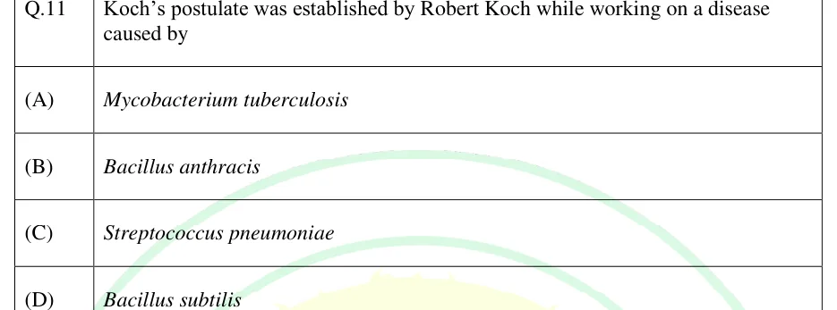
Postulates for proving that a microbe causes a disease (in order)

  1. The microbe is found in every case of the disease and not in healthy hosts.

  2. It is isolated from the diseased host and grown in pure culture.

  3. The pure culture, when inoculated into a healthy susceptible host, causes the same disease.

  4. The same microbe is re-isolated from the experimentally infected host and shown to be identical to the original.

EXAM TIP

The postulates have known exceptions. Some pathogens cannot be grown in pure culture on artificial media (obligate intracellular pathogens, for example), and some infected people carry the pathogen without disease. A question may ask which condition fails for such an organism.

Classification of prokaryotes today rests on the 16S rRNA gene (the rRNA of the 30S subunit of the 70S ribosome). It is present in all prokaryotes and performs the same function everywhere. It has conserved regions (for primer design) and variable regions (for telling groups apart). Comparing its sequences gives a phylogeny that divides life into three domains: Bacteria, Archaea and Eukarya. Phenotypic traits (shape, Gram reaction, metabolism), the G+C content of DNA, DNA-DNA hybridisation and whole-genome comparisons supplement the rRNA data.

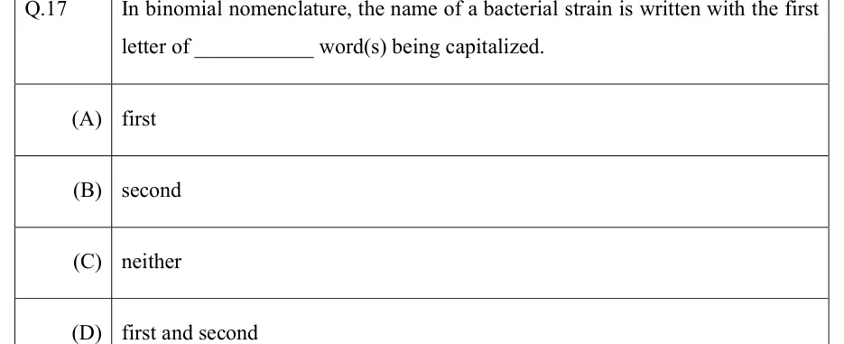
Species conventions. Two strains are conventionally placed in the same species when DNA-DNA hybridisation is about 70 percent or higher. Whole-genome average nucleotide identity (ANI) of about 95 to 96 percent is the genome-based equivalent. 16S rRNA identity below about 97 percent (a stricter modern cut-off is about 98.7 percent) indicates different species, but high 16S identity alone does not prove that two strains are the same species. Organisms are named with the binomial system: genus (capitalised) and species epithet (lower case), printed in italics, for example Escherichia coli, abbreviated E. coli. Names are written plainly on this page.

The three domains compared

Feature

Bacteria

Archaea

Eukarya

Cell type

Prokaryotic

Prokaryotic

Eukaryotic

Nuclear membrane, membrane-bound organelles

Absent

Absent

Present

Cell wall

Peptidoglycan (muramic acid present), except wall-less groups such as Mycoplasma

No peptidoglycan; S-layer, or pseudomurein in some methanogens

Absent, or chitin / cellulose; never peptidoglycan

Membrane lipids

Ester-linked fatty acids (usually unbranched) on glycerol-3-phosphate; bilayer

Ether-linked, branched isoprenoid chains on glycerol-1-phosphate; bilayer or monolayer (tetraether)

Ester-linked fatty acids on glycerol-3-phosphate; bilayer

Ribosome

70S

70S

80S (cytoplasm)

Initiator amino acid in translation

Formylmethionine

Methionine

Methionine

RNA polymerase

One enzyme with few subunits (core α2ββ'ω plus a sigma factor)

One enzyme with many subunits, resembling eukaryotic RNA polymerase II

Three nuclear enzymes (I, II, III)

Histones

Absent (histone-like nucleoid proteins instead)

Present in many

Present

Introns

Rare

Present in some tRNA and rRNA genes

Common in protein-coding genes

Methanogenesis

Absent

Present in methanogens only

Absent

Chlorophyll-based photosynthesis

Present in some (cyanobacteria, purple and green bacteria)

Absent

Present (chloroplasts)

Growth inhibited by penicillin, streptomycin, chloramphenicol

Generally yes (penicillin acts only on walled, growing cells)

No

No (cytoplasmic ribosomes and cells)

Major groups of bacteria

Group (current rank name)

Key features

Examples

Proteobacteria (Pseudomonadota)

Gram-negative; the largest and most varied phylum; classes alpha, beta, gamma and others; includes phototrophs, chemolithotrophs and many pathogens

Alpha: Rhizobium, Agrobacterium, Rickettsia. Beta: Neisseria, Nitrosomonas. Gamma: E. coli, Salmonella, Pseudomonas, Vibrio

Firmicutes (Bacillota)

Gram-positive, low G+C content; several genera form endospores

Bacillus, Clostridium, Staphylococcus, Streptococcus, Lactobacillus

Mollicutes

No cell wall; sterols in the membrane; among the smallest free-living cells; related to the low G+C Gram-positive bacteria

Mycoplasma

Actinobacteria (Actinomycetota)

Gram-positive, high G+C content; many filamentous; a major source of natural antibiotics

Streptomyces, Mycobacterium, Corynebacterium

Cyanobacteria

Oxygenic photosynthesis with chlorophyll a; many fix nitrogen, some in heterocysts

Anabaena, Nostoc, Synechococcus

Bacteroidetes (Bacteroidota)

Gram-negative rods; abundant in the gut

Bacteroides, Flavobacterium

Chlamydiae

Obligate intracellular; little or no peptidoglycan; cycle of infectious elementary bodies and dividing reticulate bodies

Chlamydia

Spirochaetes

Flexible spiral cells moved by periplasmic (axial) flagella

Treponema, Borrelia, Leptospira

Prokaryotic cell structure

Common shapes are coccus (sphere), bacillus (rod), spirillum (rigid spiral), vibrio (comma) and spirochaete (flexible spiral). Some cells, such as wall-less Mycoplasma, have no fixed shape (pleomorphic). The arrangement after division (pairs, chains, clusters) depends on the plane of division and whether cells stay attached. Prokaryotes lack a membrane-bound nucleus and membrane-bound organelles. Their chromosome lies in a region called the nucleoid, usually as one circular DNA molecule (a few bacteria, such as Borrelia and Streptomyces, have linear chromosomes).

Structures and their functions

Structure

Composition / feature

Function

Plasma membrane

Phospholipid bilayer with proteins; no sterols except in Mycoplasma (many bacteria have sterol-like hopanoids)

Selective permeability, transport, energy generation (electron transport and ATP synthase sit here)

Capsule / slime layer

Polysaccharide (sometimes polypeptide); a capsule is firmly attached, a slime layer loosely; seen by negative staining (India ink)

Protection from phagocytosis and drying, adhesion, biofilm formation

Flagellum

Filament of flagellin, hook, basal body; rotates, driven by proton motive force (sodium motive force in some, for example Vibrio). Arrangements: monotrichous (one), lophotrichous (a tuft at one end), amphitrichous (one at each end), peritrichous (all over)

Motility, including movement towards or away from chemicals (chemotaxis)

Pili / fimbriae

Protein (pilin) filaments

Attachment; the sex pilus takes part in conjugation; type IV pili give twitching motility

Endospore

Dormant, dehydrated cell with a thick coat and cortex; core rich in calcium dipicolinate and small acid-soluble proteins; seen with the endospore stain (malachite green with heat, safranin counterstain)

Survival of heat, drying, radiation and chemicals; formed by Bacillus and Clostridium

Plasmid

Small, usually circular, extra-chromosomal DNA

Carries optional genes such as antibiotic resistance

Ribosome

70S (50S + 30S subunits)

Protein synthesis; target of many antibiotics

Inclusion bodies

Glycogen, poly-beta-hydroxybutyrate, polyphosphate (volutin), sulfur granules

Storage of carbon, phosphate or sulfur

Gas vesicles; magnetosomes

Protein-walled gas bags; magnetite particles in a membrane

Buoyancy in aquatic cells; orientation along magnetic field lines

Cell walls, the Gram stain and archaea

Peptidoglycan is a single giant mesh around the cell. Its backbone alternates N-acetylglucosamine (NAG) and N-acetylmuramic acid (NAM) joined by beta-1,4 bonds. A peptide is attached to the lactyl group of each NAM. It is made as a pentapeptide: L-Ala, D-Glu, then meso-diaminopimelic acid (DAP) in E. coli and most Gram-negative bacteria (L-lysine in many Gram-positive bacteria), then D-Ala, D-Ala. In transpeptidation the terminal D-Ala is released, and the D-Ala at position 4 bonds to position 3 of a neighbouring stem, so mature stems are tetrapeptides. In most Gram-negative cells DAP bonds directly to D-Ala. In Staphylococcus aureus a pentaglycine bridge runs from L-Lys to D-Ala. Muramic acid and these D-amino acids do not occur in eukaryotic cells, which is one reason cell-wall synthesis is a good antibiotic target.

Where common agents act on the wall

  1. Lysozyme cleaves the beta-1,4 bond between NAM and NAG in the backbone.

  2. Penicillins and other beta-lactams inhibit the transpeptidases (penicillin-binding proteins), so cross-links are not formed and growing cells lyse.

  3. Vancomycin binds the D-Ala-D-Ala end of the precursor and blocks its incorporation; bacitracin blocks recycling of the lipid carrier (bactoprenol); D-cycloserine blocks the formation of D-Ala-D-Ala.

  4. Cells whose walls are removed completely are protoplasts (from Gram-positive bacteria). Partly removed walls leave spheroplasts (from Gram-negative bacteria, because the outer membrane remains). Both are osmotically fragile and survive only in an osmotically protective (isotonic or slightly hypertonic) medium.

Gram-positive and Gram-negative cell envelopes

Feature

Gram-positive

Gram-negative

Peptidoglycan

Thick, many layers (about 20 to 80 nm)

Thin, one or a few layers (a few nm)

Teichoic and lipoteichoic acids

Present

Absent

Outer membrane

Absent

Present (lipid bilayer outside the peptidoglycan, anchored to it by murein lipoprotein)

Lipopolysaccharide (LPS)

Absent

Present in the outer leaflet of the outer membrane (lipid A, core polysaccharide, O-antigen)

Porins

Absent (no outer membrane)

Present in the outer membrane (let small hydrophilic molecules pass)

Periplasmic space

Narrow or absent

Wide and prominent; holds binding proteins and enzymes

Lysozyme action

Very effective (wall is exposed)

Weak unless the outer membrane is first disturbed (for example with EDTA)

Typical susceptibility to penicillin G

Higher

Lower (the outer membrane limits entry)

Endotoxin

Absent

Present (lipid A)

Gram stain result

Purple

Pink / red

Examples

Bacillus, Staphylococcus, Clostridium

E. coli, Pseudomonas, Salmonella

Lipid A of LPS is the endotoxin: it triggers fever and shock when released from dying Gram-negative cells. The Gram stain divides most bacteria into two groups and is usually an early step in identifying an unknown isolate.

Gram stain procedure (in order, on a heat-fixed smear, with a water rinse after each reagent)

  1. Primary stain: crystal violet, which colours all cells purple.

  2. Mordant: iodine, which forms a large crystal violet-iodine complex inside the cell.

  3. Decolouriser: alcohol or acetone-alcohol.

  4. Counterstain: safranin, which colours the decolourised cells pink / red.

Mechanism. Alcohol dehydrates the thick Gram-positive peptidoglycan and shrinks its pores, so the large dye-iodine complex stays trapped and the cell remains purple. In Gram-negative cells alcohol extracts the lipid of the outer membrane, and the thin peptidoglycan cannot hold the complex. The cell is decolourised and then takes up safranin.

EXAM TIP

Over-decolourising makes Gram-positive cells look Gram-negative, and so do old cultures whose walls have broken down. Mycobacterium has a wall rich in mycolic acids (waxy lipids) and is detected by the acid-fast stain instead: carbol fuchsin driven in with heat, acid-alcohol as decolouriser (acid-fast cells stay red), and methylene blue as counterstain. Mycoplasma has no cell wall at all, so it is insensitive to penicillin and stains Gram-negative or poorly.

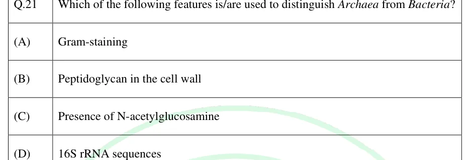
Archaea have no peptidoglycan. Their walls are an S-layer (a crystalline protein or glycoprotein sheet) or, in some methanogens, pseudomurein. Pseudomurein has N-acetyltalosaminuronic acid in place of NAM, beta-1,3 bonds in place of beta-1,4, and only L-amino acids in its peptides. Because of this, archaea resist lysozyme and penicillin. Their membrane lipids have branched isoprenoid chains joined to glycerol by ether bonds, which keeps the membranes stable at high temperature and acidity.

Well-known archaeal groups are the methanogens (strict anaerobes that make methane from H + CO or other small compounds), extreme halophiles (need about 2 to 4 M NaCl; Halobacterium uses bacteriorhodopsin, a light-driven proton pump, to make ATP) and thermoacidophiles (live in hot acidic sites, for example Sulfolobus and Thermoplasma). Methanogens and halophiles belong to the Euryarchaeota, and many hyperthermophiles to the Crenarchaeota. Many archaea also live in ordinary soils, oceans and the gut, so not all are extremophiles.

Methods in microbiology

Sterilisation and disinfection

Method

Conditions / principle

Notes

Autoclave (moist heat under pressure)

121 °C at about 15 psi (about 103 kPa) above atmospheric pressure, 15 to 20 minutes

Kills endospores; the standard for media, glassware and waste. Steam must reach all surfaces

Boiling

100 °C for about 10 minutes

Kills vegetative cells; does not reliably kill endospores, so it does not sterilise

Tyndallisation (fractional sterilisation)

Steaming at 100 °C for about 30 minutes on three successive days, with incubation between

Spores germinate between heatings and the next heating kills them; for media that cannot be autoclaved

Hot-air oven (dry heat)

160 °C for about 2 hours, or 170 °C for about 1 hour

Glassware, powders, oils; dry heat needs a higher temperature and a longer time than moist heat

Incineration / flaming

Burning; red heat of an inoculating loop

Contaminated waste, loops and needles

Pasteurisation

High temperature short time (HTST) 72 °C for 15 seconds; low temperature long time (LTLT) 63 °C for 30 minutes; ultra-high temperature (UHT) 135 to 140 °C for a few seconds

HTST and LTLT reduce pathogens and spoilage organisms but do not sterilise; UHT gives a commercially sterile product

Membrane filtration

Pore size 0.22 (0.2) micrometre retains most bacteria

For heat-sensitive solutions such as vitamins and antibiotics; Mycoplasma and viruses can pass

Radiation

Ultraviolet near 260 nm (forms thymine dimers in DNA; poor penetration); gamma rays (ionising, penetrate)

UV for surfaces and air; gamma for plastics, medical items and some foods

Ethylene oxide gas

Alkylating gas; sporicidal

Sterilises heat-sensitive plastics and medical devices

Chemical disinfectants

Alcohols, phenolics, halogens, quaternary ammonium compounds, aldehydes

Disinfectants are used on objects or surfaces, antiseptics on living tissue; most do not guarantee sterility

Killing by heat is exponential. The decimal reduction time (D-value) is the time at a fixed temperature needed to kill 90 percent of the population, that is, to lower the count by one log (base 10) unit. The z-value is the temperature rise needed to cut D to one tenth. All logarithms in this topic are base 10 unless written ln.

THERMAL DEATH (D-VALUE)

is the initial count and the survivors after time at one fixed temperature; is in the same unit as . Valid while the death curve is a straight line on a log scale.

Z-VALUE

and are the D-values at temperatures and (); is in the same temperature unit.

The F-value is the time at a reference temperature (121 °C for spores) that gives a stated kill: at that temperature. Commercial canning of low-acid foods uses a 12D process, a heating long enough to reduce spores of Clostridium botulinum by 12 log units.

Culture media

Type

Meaning

Example

Defined (synthetic)

Exact chemical composition known

Glucose-minimal salts medium

Complex

Contains digests or extracts of unknown exact composition

Nutrient broth, tryptic soy broth

Selective

Inhibits unwanted organisms and allows the wanted ones to grow

MacConkey agar (bile salts and crystal violet inhibit Gram-positive cells); mannitol salt agar (7.5 percent NaCl selects staphylococci)

Differential

Colony appearance shows a biochemical difference

MacConkey agar (lactose fermenters pink); EMB agar (E. coli colonies show a metallic green sheen); mannitol salt agar (mannitol fermenters turn the medium yellow); blood agar (haemolysis zones)

Enriched

Extra nutrients for fastidious organisms

Blood agar; chocolate agar (heated blood, for organisms such as Haemophilus and Neisseria)

Enrichment (usually liquid)

Favours the growth of one group from a mixed sample so that it becomes the majority

Selenite broth for Salmonella

Agar is a polysaccharide (agarose plus agaropectin) from red algae, used at about 1.5 percent. It melts at about 85 to 100 °C and sets at about 40 to 45 °C, and few microbes can digest it. Pure cultures (growth from a single cell, seen as a colony) are obtained by the streak-plate method (cells thinned by streaking in sectors), the spread-plate method (a small volume of diluted sample spread on a surface) or the pour-plate method (diluted sample mixed into molten agar and poured). Cultures are kept short-term in a refrigerator and long-term frozen in glycerol at about −80 °C, in liquid nitrogen, or freeze-dried (lyophilised).

Staining and microscopy. Simple stains use one dye. Differential stains (Gram, acid-fast) separate groups of cells. Special stains show capsules, spores or flagella. Total magnification is objective times eyepiece, for example with gives . Immersion oil (refractive index about 1.515, close to glass) raises NA to about 1.4, which gives a bright-field resolution limit near 0.2 micrometre. Phase-contrast and dark-field views show unstained live cells. Fluorescence microscopy uses fluorescent dyes or labelled antibodies. Electron microscopes resolve far finer detail (TEM for internal structure, SEM for surfaces).

RESOLVING POWER OF A LIGHT MICROSCOPE

is the smallest resolvable distance, the wavelength, the refractive index between specimen and objective and half the angle of the cone of light entering the objective. This is the Rayleigh form; some texts use . A smaller means better resolution.

Counting cells. A direct count under the microscope with a counting chamber (haemocytometer) includes dead cells, and so do electronic counters and turbidity (optical density) readings, which give a quick measure of biomass. A viable count counts only cells able to form colonies. Because a colony may arise from a single cell or from a clump, the result is reported as colony-forming units (CFU). Choose a plate with 30 to 300 colonies (some standards use 25 to 250). Membrane-filter counts suit dilute samples such as water. The most probable number (MPN) method estimates viable numbers statistically from growth in replicate tubes of a dilution series.

VIABLE COUNT FROM SERIAL DILUTION

Here the dilution factor is the fraction left after all dilutions, for example after six tenfold steps. Equivalently, CFU/mL = colonies × reciprocal of the dilution ÷ volume plated.

WORKED EXAMPLE

Spreading 0.1 mL of a dilution of a culture gave 85 colonies. Find the viable count of the original culture in CFU/mL.

Solution
  1. The count of 85 lies in the reliable range of 30 to 300.

  2. CFU/mL .

  3. .

Answer: CFU/mL

Microbial growth

Bacteria multiply by binary fission: one cell grows, replicates its DNA and divides into two. In balanced exponential growth the number doubles at a constant interval, the generation time (doubling time) . After generations, .

Phases of a batch-culture growth curve

Phase

What happens

Plot of log N

Lag

Cells adapt to the new medium (make enzymes, repair damage); they may grow in size but there is no net increase in number. Longer for old inocula or a changed medium

Flat

Exponential (log)

Fastest growth, constant generation time, cells most uniform

Straight line with positive slope

Stationary

Nutrient depletion or toxic wastes stop net growth; the number of viable cells stays constant; secondary metabolites and endospores often appear

Flat

Death (decline)

Viable count falls, usually exponentially

Straight line with negative slope

NUMBER OF GENERATIONS

Use only for the exponential phase. and are the cell counts at the start and end of the interval.

GENERATION TIME AND DIVISION RATE CONSTANT

is the elapsed exponential time; is generations per unit time.

SPECIFIC GROWTH RATE

Per unit time (for example per hour). It uses the natural log, so .

WORKED EXAMPLE

A culture in exponential growth rises from to cells/mL in 5 hours. Find the generation time.

Solution
  1. .

  2. generations.

  3. h min, so min.

Answer: About 31 minutes ( generations)

EXAM TIP

Diauxic growth. In a medium with glucose and lactose, E. coli uses glucose first. When glucose runs out there is a second, short lag while the lactose-utilising enzymes are induced, then a second exponential phase on lactose. The cause is catabolite repression of the lac operon by glucose.

Batch and continuous culture. A batch culture is a closed system: nutrients fall and wastes build up, giving the four phases above. A chemostat is an open system. Fresh medium with limiting-nutrient concentration flows in at rate , culture leaves at the same rate, and the volume is constant. The dilution rate is (not to be confused with the D-value). At steady state, cell density and substrate concentration are constant and the specific growth rate equals the dilution rate (). Cells wash out if exceeds the critical value , which is slightly below . A turbidostat instead keeps turbidity constant by adjusting the flow.

MONOD GROWTH EQUATION

is the limiting nutrient concentration; is the value of at which (units of concentration). It applies when one nutrient is limiting.

CHEMOSTAT STEADY STATE

and are the steady-state substrate and biomass concentrations, and is the biomass yield (mass of cells formed per mass of substrate used). This assumes Monod kinetics with no cell death and no maintenance consumption.

Environmental factors and growth classes

Factor

Classes

Temperature (optimum)

Psychrophile below about 15 °C (cannot grow above about 20 °C); psychrotroph grows at 0 °C but has its optimum at about 20 to 30 °C; mesophile about 20 to 45 °C; thermophile about 45 to 80 °C; hyperthermophile above about 80 °C

pH (optimum)

Acidophile below about pH 5.5; neutrophile about pH 5.5 to 8; alkaliphile above about pH 8

Oxygen

Obligate aerobe (needs O); microaerophile (needs low O, harmed by air levels); facultative anaerobe (grows with or without O, better with); aerotolerant anaerobe (indifferent to O, ferments only); obligate anaerobe (killed or inhibited by O)

Solutes and water

Halophiles need NaCl (extreme halophiles about 2 to 4 M); halotolerant cells grow with or without salt; osmophiles tolerate high sugar; most bacteria need a high water activity (about 0.9 or more). Walled cells resist bursting in hypotonic medium because the wall withstands the turgor; wall-less cells (protoplasts, Mycoplasma) burst. All cells plasmolyse (shrink) in hypertonic medium, which is why salt and sugar preserve food

EXAM TIP

Obligate aerobes and facultative anaerobes make both superoxide dismutase (SOD) and catalase to remove toxic oxygen products. Aerotolerant anaerobes have SOD but no catalase (some use a peroxidase). Microaerophiles have low levels of these enzymes. Obligate anaerobes lack both or have very little, which is why oxygen kills them. In a thioglycollate tube, obligate aerobes grow at the top, obligate anaerobes at the bottom, facultative anaerobes throughout but densest at the top, aerotolerant anaerobes evenly throughout, and microaerophiles in a band just below the surface.

Microbial nutrition

All cells need the macronutrients carbon, hydrogen, oxygen, nitrogen, sulfur and phosphorus, together with potassium, magnesium, calcium and iron. They also need trace elements (micronutrients) such as zinc, manganese, cobalt, molybdenum, nickel and copper, often as enzyme cofactors. A growth factor is an organic compound that the cell needs but cannot make itself, for example a vitamin, an amino acid, or a purine or pyrimidine. A mutant that needs such a compound is an auxotroph. A strain that makes it, usually the wild type, is a prototroph.

Nutritional types

Type

Energy source

Carbon source

Examples

Photoautotroph

Light

CO

Cyanobacteria, purple and green sulfur bacteria, plants, algae

Photoheterotroph

Light

Organic compounds

Purple non-sulfur and green non-sulfur bacteria

Chemolithoautotroph

Oxidation of inorganic compounds (NH, NO, HS, S, Fe, H)

CO

Nitrifying bacteria, sulfur-oxidising bacteria, iron-oxidising bacteria, hydrogen bacteria, many methanogens

Chemoheterotroph (chemoorganoheterotroph)

Oxidation of organic compounds

Organic compounds

E. coli, animals, fungi, most pathogens

Phototroph or chemotroph describes the energy source. Lithotroph or organotroph describes the electron source (inorganic or organic). Autotroph or heterotroph describes the carbon source (CO or organic compounds). Full names combine all three, for example chemolithoautotroph. Cyanobacteria carry out oxygenic photosynthesis: they use water as the electron donor, have chlorophyll a and two photosystems, and release O. Purple and green bacteria carry out anoxygenic photosynthesis: they use HS, H or organic compounds as electron donors, have bacteriochlorophylls and one photosystem, and release no O.

Nutrient uptake across the membrane

Mechanism

Energy

Feature

Simple diffusion

None

Small uncharged molecules and gases; down a concentration gradient

Facilitated diffusion

None

Carrier or channel protein; down the gradient; the substance is unchanged

Primary active transport

ATP (for example ABC transporters)

Against the gradient; uses periplasmic binding proteins in Gram-negative cells (membrane-anchored binding lipoproteins in Gram-positive cells)

Secondary active transport

Proton motive force or sodium gradient

Symport or antiport (for example lactose permease, a lactose-H symporter)

Group translocation

Phosphoenolpyruvate

The substrate is chemically changed in transit (the phosphotransferase system phosphorylates sugars such as glucose)

Siderophore uptake

Proton motive force (through TonB) at the outer-membrane receptor in Gram-negative cells, then ATP at an inner-membrane ABC transporter

Secreted iron-chelating molecules bind Fe and bring it into the cell

EXAM TIP

In group translocation the molecule inside the cell is not the same as the molecule outside (it is phosphorylated), so it is the only mechanism in the table where the transported substance is chemically modified.

PREVIOUS-YEAR QUESTIONS

8 questions from the BT papers

Try each question first, then open the answer. We placed each question under a topic ourselves; the exam paper does not label topics.

See these papers in full: GATE BT 2026 · GATE BT 2025 · GATE BT 2024 · GATE BT 2023 · GATE BT 2022 · GATE BT 2020

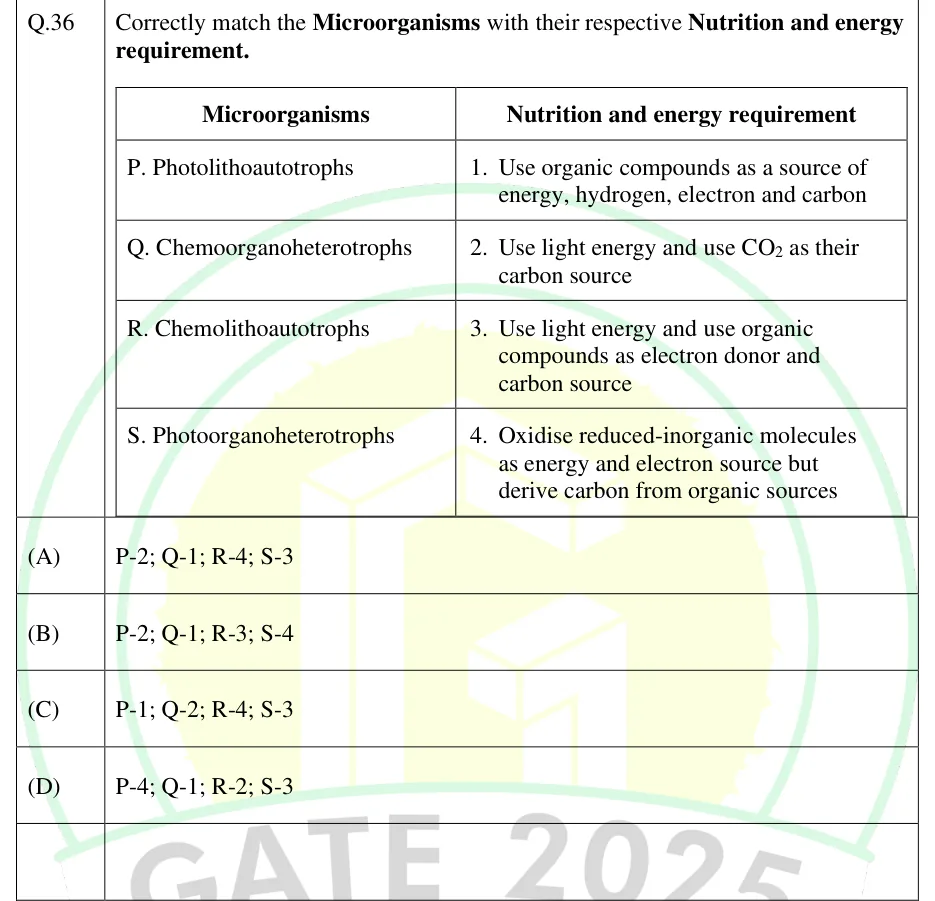
GATE 2025 BT Q.36

GATE 2025 · BTQ.36 · 2 marks · MCQ

Correctly match the Microorganisms with their respective Nutrition and energy requirement.

Microorganisms: P. Photolithoautotrophs; Q. Chemoorganoheterotrophs; R. Chemolithoautotrophs; S. Photoorganoheterotrophs.

Nutrition and energy requirement: 1. Use organic compounds as a source of energy, hydrogen, electron and carbon; 2. Use light energy and use CO2 as their carbon source; 3. Use light energy and use organic compounds as electron donor and carbon source; 4. Oxidise reduced-inorganic molecules as energy and electron source but derive carbon from organic sources.

GATE 2025 BT question 36: A two-column table.Tap to open full size
Figure from the GATE 2025 BT paper. It is part of the question.
Figure description

A two-column table. Left column 'Microorganisms' with rows P, Q, R, S as above. Right column 'Nutrition and energy requirement' with rows 1 to 4 as above.

  1. (A)

    P-2; Q-1; R-4; S-3

  2. (B)

    P-2; Q-1; R-3; S-4

  3. (C)

    P-1; Q-2; R-4; S-3

  4. (D)

    P-4; Q-1; R-2; S-3

Answer and solution
GATE 2025 BT Q.36: answer and solution

GATE 2023 BT Q.37

GATE 2023 · BTQ.37 · 2 marks · MCQ

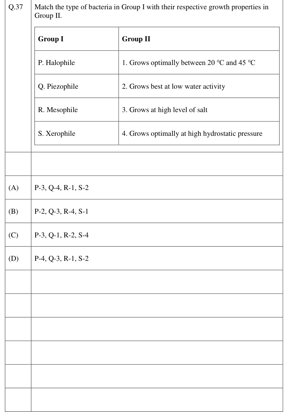
Match the type of bacteria in Group I with their respective growth properties in Group II.

Group I: P. Halophile; Q. Piezophile; R. Mesophile; S. Xerophile.
Group II: 1. Grows optimally between 20 °C and 45 °C; 2. Grows best at low water activity; 3. Grows at high level of salt; 4. Grows optimally at high hydrostatic pressure.

GATE 2023 BT question 37: A two-column matching table.Tap to open full size
Figure from the GATE 2023 BT paper. It is part of the question.
Figure description

A two-column matching table. Header row: Group I | Group II. Rows: P. Halophile | 1. Grows optimally between 20 °C and 45 °C; Q. Piezophile | 2. Grows best at low water activity; R. Mesophile | 3. Grows at high level of salt; S. Xerophile | 4. Grows optimally at high hydrostatic pressure.

  1. (A)

    P-3, Q-4, R-1, S-2

  2. (B)

    P-2, Q-3, R-4, S-1

  3. (C)

    P-3, Q-1, R-2, S-4

  4. (D)

    P-4, Q-3, R-1, S-2

Answer and solution
GATE 2023 BT Q.37: answer and solution

GATE 2020 BT Q.16

GATE 2020 · BTQ.16 · 1 mark · MCQ

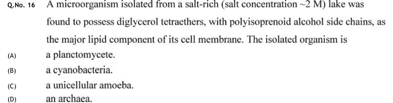
A microorganism isolated from a salt-rich (salt concentration ~2 M) lake was found to possess diglycerol tetraethers, with polyisoprenoid alcohol side chains, as the major lipid component of its cell membrane. The isolated organism is

  1. (A)

    a planctomycete.

  2. (B)

    a cyanobacteria.

  3. (C)

    a unicellular amoeba.

  4. (D)

    an archaea.

Answer and solution
See the question in the paper
GATE 2020 BT Q.16: answer and solution

PRACTICE

6 questions to test yourself

PRACTICE 11 mark · MCQ

Which of the following is characteristic of Gram-negative bacteria and absent from Gram-positive bacteria?

  1. (A)

    Lipoteichoic acid anchored in the plasma membrane

  2. (B)

    Lipopolysaccharide in the outer leaflet of an outer membrane

  3. (C)

    A thick multilayered peptidoglycan with a pentaglycine cross-bridge

  4. (D)

    Pseudomurein as the main wall polymer

Answer and solution

Answer: (B)

  1. Lipoteichoic acid is a Gram-positive feature, so A is wrong.

  2. A thick peptidoglycan with a pentaglycine bridge (as in Staphylococcus aureus) is also Gram-positive, so C is wrong.

  3. Pseudomurein occurs in some archaea, not in Gram-negative bacteria, so D is wrong.

  4. Gram-negative cells have an outer membrane whose outer leaflet carries LPS, so B is correct.

PRACTICE 22 marks · NAT

0.2 mL of a dilution of a bacterial culture was spread on an agar plate and gave 150 colonies. Calculate the viable count of the original culture. Give the answer in units of CFU/mL.

Answer and solution

Answer: (accepted range )

  1. CFU/mL = colonies / (volume plated × dilution factor).

  2. .

  3. CFU/mL, so the answer is 7.5 in units of .

PRACTICE 32 marks · NAT

A bacterial culture in exponential growth increases from cells/mL to cells/mL in 4 hours. What is the generation time in minutes? Give the answer to one decimal place.

Answer and solution

Answer: (accepted range )

  1. .

  2. generations.

  3. h min, so min, about 24.1 min.

PRACTICE 42 marks · MSQ

Which of the following statements about Archaea are correct?

  1. (A)

    Their membrane lipids contain ether-linked isoprenoid chains

  2. (B)

    Their cell walls contain peptidoglycan with N-acetylmuramic acid

  3. (C)

    Their cells are not digested by lysozyme because they lack peptidoglycan

  4. (D)

    Translation starts with methionine, not formylmethionine

Answer and solution

Answer: (A), (C), (D)

  1. A is correct: archaeal lipids are ether-linked and isoprenoid.

  2. B is wrong: archaea have no peptidoglycan; their walls are S-layers or pseudomurein.

  3. C is correct: lysozyme cuts the beta-1,4 NAM-NAG bond of peptidoglycan, which archaea do not have.

  4. D is correct: archaea, like eukaryotes, use methionine as the initiator amino acid.

PRACTICE 51 mark · MCQ

A vial contains spores of a heat-resistant bacterium. At 121 °C the D-value of these spores is 1.5 min. For how long must the vial be held at 121 °C so that the expected number of surviving spores per vial falls to ?

  1. (A)

    6 min

  2. (B)

    9 min

  3. (C)

    12 min

  4. (D)

    15 min

Answer and solution

Answer: (C)

  1. .

  2. min.

  3. So option C is correct.

PRACTICE 62 marks · NAT

A chemostat is fed with medium containing 10 g/L of the growth-limiting substrate. The organism follows Monod kinetics with h and g/L, and its biomass yield is g cells per g substrate. The dilution rate is 0.25 h. Assuming no cell death and no maintenance consumption, what is the steady-state biomass concentration in g/L? Give the answer to one decimal place.

Answer and solution

Answer: (accepted range )

  1. At steady state h, which is below the washout limit h.

  2. g/L.

  3. g/L.

OFFICIAL GATE 2027 SYLLABUS WORDING

Copied from the GATE 2027 syllabus; see all 2027 syllabi.

GATE BT

Microbiology: History of Microbiology, Bacterial classification and diversity; Bacterial cell wall composition, Gram positive and Gram-negative bacteria, Archea, Methods in microbiology; Microbial growth and nutrition

Preparing in Kolkata? IAE Academy's course for GATE BT.