GATE 2025 BT Q.15
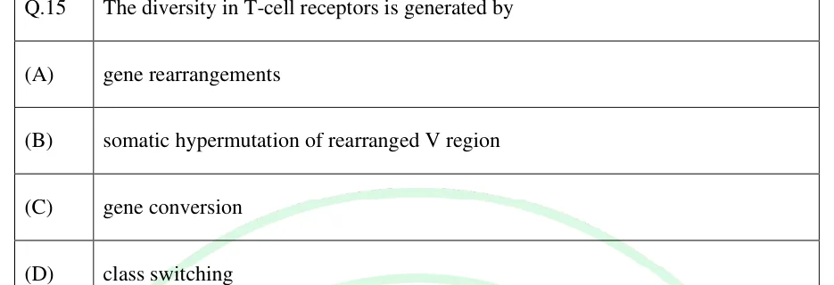
The diversity in T-cell receptors is generated by
- (A)
gene rearrangements
- (B)
somatic hypermutation of rearranged V region
- (C)
gene conversion
- (D)
class switching

GATE BT
T and B cells, MHC, antigen presentation and immune regulation
This topic covers how T and B lymphocytes develop and are selected, how MHC molecules present peptides to T cells, and how the immune response is switched on, remembered, limited and sometimes misdirected (hypersensitivity, autoimmunity, immunodeficiency, graft rejection). It also covers how vaccines exploit memory. GATE questions test the logic linking receptor, MHC class, co-receptor, pathway and outcome, so learning the comparison tables makes the questions quick to solve.
7 previous-year questions · 7 solved · 5 practice questions
Both T and B cells arise from haematopoietic stem cells. B cells mature in the bone marrow. T-cell precursors travel to the thymus. In both organs the antigen receptor genes are assembled by V(D)J recombination, started by the recombinase enzymes RAG1 and RAG2 and completed by DNA-repair enzymes, and the cells are then tested for useful and harmless receptors.
T-cell development in the thymus, in order
Double-negative (DN, CD4⁻CD8⁻) thymocytes rearrange the TCR β chain and display it with the pre-Tα chain as the pre-TCR. A minority rearrange γ and δ chains instead and become γδ T cells, which mostly do not need classical MHC presentation.
Pre-TCR signalling stops further β rearrangement (allelic exclusion), and cells proliferate, become double-positive (DP, CD4⁺CD8⁺) and rearrange the α chain.
Positive selection (thymic cortex, cortical epithelial cells): a TCR that binds self-MHC with low-to-moderate affinity receives a survival signal. Cells that cannot bind self-MHC die by neglect. This creates MHC restriction.
Lineage commitment: a DP cell whose TCR recognises MHC class I becomes a single-positive CD8⁺ cell, and one that recognises MHC class II becomes a single-positive CD4⁺ cell.
Negative selection (cortico-medullary junction and medulla, medullary epithelial cells and dendritic cells): strong binding to self-peptide–MHC causes apoptosis (clonal deletion). The transcription factor AIRE makes medullary epithelial cells express many tissue-restricted self-antigens; AIRE mutation causes APECED (autoimmune polyendocrine syndrome type 1). Some self-reactive CD4 cells are diverted to become regulatory T cells.
B-cell development is ordered as follows. The heavy chain is rearranged first and appears with the surrogate light chain (VpreB and λ5) as the pre-BCR. Pre-BCR signalling stops rearrangement at the other heavy-chain allele (allelic exclusion), so each B cell makes receptors of one specificity. The light chain is then rearranged (κ usually before λ), giving an immature IgM⁺ B cell. Immature cells that bind self-antigen strongly in the marrow undergo receptor editing (a new light-chain rearrangement), clonal deletion or anergy. Surviving cells leave for the spleen and lymph nodes and become mature naive B cells that carry IgM and IgD.
T-cell subsets
Subset | Key transcription factor | Signature cytokines or effector molecules | Main function |
|---|---|---|---|
Th1 (CD4) | T-bet | IFN-γ (induced by IL-12) | Activates macrophages, defence against intracellular pathogens |
Th2 (CD4) | GATA3 | IL-4, IL-5, IL-13 (induced by IL-4) | Helminths, IgE class switching, eosinophil activation; allergy |
Th17 (CD4) | RORγt | IL-17, IL-22 (induced by TGF-β with IL-6) | Neutrophil recruitment, extracellular bacteria and fungi |
Tfh (CD4) | Bcl6 | IL-21; CXCR5 homing to follicles | Help to B cells in germinal centres |
Treg (CD4) | FOXP3 (CD25⁺) | IL-10, TGF-β | Suppress immune responses, maintain tolerance |
CD8 cytotoxic (CTL) | Eomes, T-bet | IFN-γ; perforin and granzymes | Kill virus-infected and tumour cells (also by Fas–FasL) |
EXAM TIP
A naive T cell needs two signals to be activated: signal 1 is TCR binding to peptide–MHC (with CD4 or CD8), and signal 2 is co-stimulation, mainly CD28 on the T cell binding B7 (CD80/CD86) on the antigen-presenting cell. Cytokines that steer differentiation are often called signal 3. Signal 1 without co-stimulation makes a naive T cell anergic.
The MHC is a cluster of closely linked genes. In humans it is called HLA and lies on chromosome 6. In mouse it is H-2 on chromosome 17. Its products present peptides to T cells, and they are the main targets of graft rejection. Class I and class II genes are highly polymorphic.
MHC class I versus class II
Feature | Class I | Class II |
|---|---|---|
Human genes | HLA-A, B, C | HLA-DR, DP, DQ |
Mouse genes | H-2K, D, L | I-A, I-E |
Chains | One heavy α chain (α1, α2, α3) plus β2-microglobulin (not MHC-encoded, not polymorphic) | α chain and β chain (two domains each, both MHC-encoded) |
Peptide-binding groove | Formed by α1 and α2; closed ends | Formed by α1 and β1; open ends |
Peptide length | About 8 to 10 amino acids | About 13 to 25 amino acids |
Co-receptor binding | CD8 binds α3 | CD4 binds β2 |
Presents to | CD8⁺ T cells | CD4⁺ T cells |
Expressed on | All nucleated cells (levels vary between tissues; absent from mature red cells) | Dendritic cells, macrophages, B cells, thymic epithelium (and activated human T cells); IFN-γ can induce it on other cells such as endothelium |
Source of peptides | Cytosolic (endogenous) proteins | Endocytosed (exogenous) proteins |
Class III genes lie between class I and II. They code for proteins that do not present antigen, including complement components C2, C4 and factor B, and the cytokines TNF and lymphotoxin.
MHC diversity in a population comes from polygeny (several class I and class II loci) and extreme polymorphism (many alleles per locus), concentrated in the residues that form the peptide groove. Alleles are codominant: a person expresses both parental alleles. A heterozygote therefore has up to six different classical class I molecules (two each of A, B and C), and at least six classical class II molecules (DR, DP and DQ from each parent), often more because some haplotypes carry an extra DRB gene and DQ and DP α and β chains from the two parents can pair in mixed combinations. The set of MHC genes on one chromosome is a haplotype and is usually inherited as a block, because recombination within the complex is rare. Population diversity makes it unlikely that a pathogen can escape presentation in every member of the population. A T cell recognises its peptide only when it is bound to the self-MHC allele it was selected on (MHC restriction).
HLA MATCH BETWEEN TWO FULL SIBLINGS
Applies when the two parents carry four different haplotypes between them and there is no recombination within the MHC. Each child receives one of two maternal and one of two paternal haplotypes, each with probability 1/2.
EXAM TIP
β2-microglobulin is encoded outside the MHC (chromosome 15 in humans). Its absence prevents class I from reaching the cell surface. NK cells attack cells that have lost class I ('missing self').
Cytosolic (endogenous) pathway to MHC class I
Cytosolic proteins, including viral proteins, are cut by the proteasome (the immunoproteasome, with LMP2 and LMP7, is induced by IFN-γ; the LMP2 and LMP7 genes lie in the MHC).
TAP1/TAP2, also encoded in the MHC, transports peptides into the ER lumen. ERAP can trim them.
In the ER, the newly made class I heavy chain is held by calnexin until β2-microglobulin binds. The heavy chain–β2-microglobulin dimer then joins the peptide-loading complex (tapasin, calreticulin, ERp57, TAP). A peptide binds and stabilises the complex.
The peptide–class I complex travels through the Golgi to the cell surface and is shown to CD8⁺ T cells.
Endocytic (exogenous) pathway to MHC class II
Antigen is taken up by phagocytosis, endocytosis or the B-cell receptor and is degraded by acid proteases (cathepsins) in endosomes.
Class II α and β chains are made in the ER with the invariant chain (Ii), which blocks the groove and directs the complex to the endocytic compartment (MIIC).
Ii is degraded, leaving a small fragment called CLIP in the groove.
HLA-DM (H-2M in mouse) exchanges CLIP for an antigenic peptide. HLA-DO inhibits HLA-DM, mainly in B cells.
The peptide–class II complex is displayed to CD4⁺ T cells.
Cross-presentation is the loading of exogenous antigen onto MHC class I. It is carried out especially by a subset of dendritic cells and primes CD8⁺ T cells against viruses that do not infect dendritic cells and against tumours. Professional antigen-presenting cells (dendritic cells, macrophages, B cells) express class II and co-stimulatory molecules. Dendritic cells are the most effective at activating naive T cells.
EXAM TIP
Superantigens (such as staphylococcal enterotoxins and toxic shock syndrome toxin) bind outside the peptide groove, linking MHC class II to the Vβ region of many TCRs. They activate a large fraction of T cells irrespective of antigen specificity and cause massive cytokine release.
T-dependent versus T-independent responses
Feature | T-dependent (protein antigens) | T-independent (TI-1 and TI-2) |
|---|---|---|
Examples | Tetanus toxoid, viral proteins | TI-1: LPS; TI-2: repeating polysaccharides such as pneumococcal capsule |
T-cell help | Required (Tfh, CD40L–CD40, cytokines) | Not required |
Mechanism | BCR binds antigen, B cell presents peptide on class II to Tfh | TI-1: mitogenic (polyclonal at high dose) signal; TI-2: extensive BCR cross-linking |
Germinal centre, somatic hypermutation, affinity maturation | Yes | Little or none |
Class switching | Yes (IgG, IgA, IgE) | Limited; mostly IgM |
Memory | Yes | Poor |
In a germinal centre, activation-induced cytidine deaminase (AID) drives both somatic hypermutation of the V genes and class-switch recombination of the constant region. B cells whose mutated receptors bind antigen better are selected, which is affinity maturation. Cells leave as long-lived plasma cells or memory B cells. T-cell cytokines steer the switch: IL-4 favours IgE, TGF-β favours IgA, and IFN-γ favours opsonising IgG subclasses (IgG2a in mice). Absence of CD40 ligand (X-linked) causes hyper-IgM syndrome: IgM is normal or raised but switched isotypes are deficient. Defects in CD40 or AID cause other forms of hyper-IgM syndrome.
Primary versus secondary response to a protein (T-dependent) antigen
Feature | Primary response | Secondary (memory) response |
|---|---|---|
Lag phase | Longer (several days) | Shorter |
Peak antibody level | Lower | Higher |
Main isotype | IgM first, then IgG | IgG predominates (also IgA, IgE) |
Affinity | Lower | Higher |
Cells responding | Naive lymphocytes | Memory B and T cells, more numerous and with a lower activation threshold |
Memory T cells include central memory cells (CCR7⁺, home to lymph nodes) and effector memory cells (CCR7⁻, found in blood and peripheral tissues); tissue-resident memory cells stay in one tissue. Memory persists after antigen is cleared and is the basis of immunisation.
An immune response must stop when the antigen is cleared and must not attack self. Regulation works through several mechanisms.
Mechanisms of regulation
Regulatory T cells (FOXP3⁺, CD25⁺) secrete IL-10 and TGF-β and consume IL-2. FOXP3 mutation causes IPEX, a severe autoimmune disease.
CTLA-4 on activated T cells and Tregs binds B7 with higher affinity than CD28, delivers an inhibitory signal and removes B7 from antigen-presenting cells. PD-1 is another inhibitory receptor (ligands PD-L1 and PD-L2); blocking CTLA-4 or PD-1 is used in cancer immunotherapy.
Activation-induced cell death: repeated stimulation raises Fas and FasL, so activated T cells die by apoptosis. Fas mutations cause autoimmune lymphoproliferative syndrome (ALPS).
Antibody feedback: IgG–antigen complexes bind FcγRIIB on B cells and switch them off.
Cytokines such as IL-10 and TGF-β suppress macrophage and T-cell function.
Central versus peripheral tolerance
Feature | Central tolerance | Peripheral tolerance |
|---|---|---|
Site | Thymus (T cells), bone marrow (B cells) | Tissues and secondary lymphoid organs |
Mechanisms | Clonal deletion, receptor editing (B cells), Treg diversion | Anergy (signal 1 without co-stimulation), deletion, Treg suppression, ignorance (including antigens in immune-privileged sites) |
Limit | Only antigens present in the primary organs are encountered (aided by AIRE) | Catches self-reactive cells that escaped |
Hypersensitivity
Type | Effector | Mechanism | Examples |
|---|---|---|---|
I (immediate) | IgE, mast cells and basophils | Allergen cross-links IgE on FcεRI, releasing histamine and other mediators within minutes; a late-phase reaction can follow hours later | Anaphylaxis, allergic rhinitis, atopic asthma |
II (antibody against cell or matrix antigens) | IgG or IgM, complement, phagocytes, NK cells | Opsonisation, complement lysis, ADCC, or altered receptor function (receptor-stimulating or blocking antibodies are sometimes called type V) | ABO-incompatible transfusion reaction, haemolytic disease of the newborn, Goodpasture syndrome, myasthenia gravis, Graves disease |
III (immune complex) | Soluble antigen–antibody complexes | Deposition in vessels and tissues, complement activation, neutrophil damage | Serum sickness, Arthus reaction, SLE, post-streptococcal glomerulonephritis |
IV (delayed type) | T cells (Th1, CD8) and macrophages | Cytokine release and cell killing, peak after 24 to 72 hours | Contact dermatitis, tuberculin test, granuloma formation |
Autoimmunity is an immune response against self and may be organ-specific (Hashimoto thyroiditis, type 1 diabetes, multiple sclerosis) or systemic (SLE, rheumatoid arthritis). Contributing factors include failure of tolerance, molecular mimicry, release of sequestered antigens, certain HLA alleles and inflammation that provides co-stimulation.
Standard HLA associations with disease
HLA allele | Associated disease |
|---|---|
HLA-B27 | Ankylosing spondylitis |
HLA-DR3, DR4 (with DQ2, DQ8) | Type 1 diabetes |
HLA-DR4 | Rheumatoid arthritis |
HLA-DQ2, DQ8 | Coeliac disease |
Immunodeficiency
Condition | Defect | Consequence |
|---|---|---|
SCID | Examples: ADA deficiency, RAG1/2 mutation, IL2RG (common γ chain, X-linked), JAK3 | No functional T cells, so antibody responses fail even where B cells are present (B cells are also absent in RAG and ADA deficiency); severe infections from infancy |
X-linked agammaglobulinaemia | BTK mutation, B cells arrest at the pre-B stage | Very low immunoglobulins, recurrent bacterial infections |
DiGeorge syndrome | Thymic hypoplasia (22q11.2 deletion), with parathyroid and heart defects | Low T cells, low calcium |
Chronic granulomatous disease | NADPH oxidase defect | Poor killing of ingested microbes, granulomas |
Hyper-IgM syndrome | CD40L (X-linked), CD40 or AID defect | No class switching |
Late complement deficiency | C5 to C9 | Recurrent Neisseria infections |
C1-inhibitor deficiency | Uncontrolled complement and kinin activation | Hereditary angioedema |
AIDS (acquired) | HIV | Loss of CD4⁺ T cells |
HIV is a retrovirus (a lentivirus). Its envelope glycoprotein gp120 binds CD4 and a chemokine co-receptor (CCR5 or CXCR4), and gp41 mediates membrane fusion. Reverse transcriptase makes DNA, integrase inserts it into the host genome, and CD4⁺ T cells, macrophages and dendritic cells are infected. People homozygous for the CCR5-Δ32 deletion are highly resistant to CCR5-using strains. AIDS is defined clinically by opportunistic infections or CD4⁺ counts below 200 cells per microlitre.
In transplantation, a graft from one site to another in the same individual is an autograft, and one between genetically identical individuals is an isograft. Between members of one species it is an allograft, and between species a xenograft. Rejection is driven mainly by differences in MHC, with minor histocompatibility antigens contributing; ABO antigens matter for hyperacute rejection.
Hyperacute rejection (minutes to hours) is due to preformed antibodies against ABO or donor HLA that activate complement on the graft endothelium. Acute rejection (days to weeks) is mainly T-cell mediated, although antibody-mediated acute rejection also occurs. Chronic rejection develops over months to years, with thickening of graft vessels and fibrosis. Cyclosporin A (bound to cyclophilin) and tacrolimus (bound to FKBP12) block calcineurin, so NFAT is not activated and IL-2 gene transcription falls. Rapamycin (bound to FKBP12) inhibits mTOR and so blocks the T-cell proliferation that IL-2 drives.
Graft-versus-host disease (GVHD) occurs when immunocompetent donor T cells in a graft, typically a bone marrow or haematopoietic stem cell graft, attack the tissues of an immunocompromised recipient, especially skin, liver and gut. It needs three conditions: immunocompetent donor cells, host antigens foreign to the donor, and a host unable to reject the donor cells. HLA matching and T-cell depletion of the graft reduce it. The same donor T cells can also attack residual leukaemia cells (graft-versus-leukaemia effect).
Active immunisation gives antigen so the person makes their own response and memory. Passive immunisation gives ready-made antibody (antitoxin, immunoglobulin, maternal IgG across the placenta, anti-D immunoglobulin to prevent Rh haemolytic disease of the newborn). It acts at once but lasts only weeks to a few months and does not create memory.
Vaccine types
Type | Principle | Examples | Points |
|---|---|---|---|
Live attenuated | Weakened replicating organism | BCG, oral polio (Sabin), measles, mumps, rubella, varicella, yellow fever | Strong humoral and cellular response, long-lasting; may revert; avoided in immunodeficiency and pregnancy |
Inactivated (killed) | Whole organism killed | Injected polio (Salk), rabies, hepatitis A | Safer, mainly humoral; boosters needed |
Subunit / recombinant | Purified or recombinant antigen | Hepatitis B (recombinant surface antigen made in yeast), HPV (virus-like particles) | No infection risk; usually needs adjuvant |
Toxoid | Inactivated toxin | Tetanus, diphtheria | Induces anti-toxin antibody |
Conjugate | Polysaccharide linked to protein carrier | Hib, pneumococcal conjugate, meningococcal conjugate | Converts a T-independent antigen into a T-dependent one, so infants under about 2 years form memory |
mRNA | Lipid-nanoparticle-delivered mRNA for an antigen | SARS-CoV-2 mRNA vaccines | Host cells make the antigen; cellular and humoral responses |
Viral vector | Harmless virus carrying an antigen gene | Adenovirus-vector vaccines | Cellular and humoral responses |
Adjuvants such as aluminium salts enhance the response by activating innate immunity. Herd immunity protects those unvaccinated when enough of the population is immune that each case infects fewer than one other person on average.
VACCINATION COVERAGE NEEDED FOR HERD IMMUNITY
is the basic reproduction number and is vaccine efficacy (fraction, 0 to 1). is the herd immunity threshold, the coverage needed when . Assumes homogeneous mixing, and applies when is high enough that does not exceed 1.
WORKED EXAMPLE
A parent pair has HLA haplotypes a/b (mother) and c/d (father), all different. A patient is a/c. What is the probability that a new full sibling is HLA-identical to the patient, and that a new sibling shares no haplotype?
Each child gets one maternal haplotype (a or b) and one paternal (c or d), each with probability 1/2.
Identical to the patient needs a from the mother and c from the father: 1/2 × 1/2 = 1/4.
No shared haplotype needs b and d: 1/2 × 1/2 = 1/4.
Answer: 1/4 for an identical sibling, and 1/4 for a sibling sharing none (1/2 for sharing exactly one).
WORKED EXAMPLE
A pathogen has and a vaccine has efficacy 0.8. What fraction of the population must be vaccinated for herd immunity?
Immune fraction needed = 1 − 1/4 = 0.75.
Coverage = 0.75 / 0.8 = 0.9375.
Answer: About 93.75 percent.
PREVIOUS-YEAR QUESTIONS
Try each question first, then open the answer. We placed each question under a topic ourselves; the exam paper does not label topics.
See these papers in full: GATE BT 2025 · GATE BT 2023 · GATE BT 2020
The diversity in T-cell receptors is generated by
gene rearrangements
somatic hypermutation of rearranged V region
gene conversion
class switching
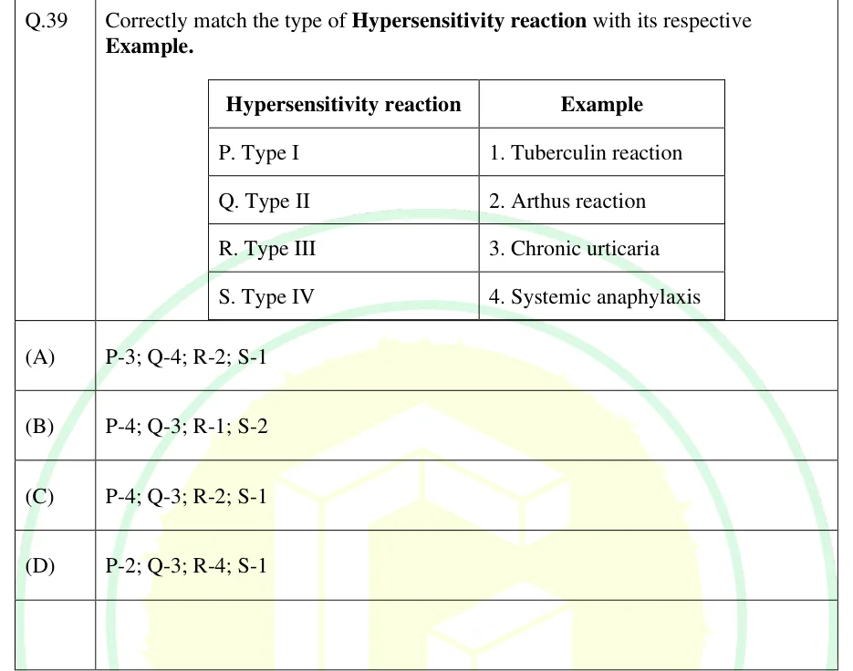
Correctly match the type of Hypersensitivity reaction with its respective Example.
 Tap to open full size
A two-column table. Header: Hypersensitivity reaction | Example. Rows: P. Type I | 1. Tuberculin reaction; Q. Type II | 2. Arthus reaction; R. Type III | 3. Chronic urticaria; S. Type IV | 4. Systemic anaphylaxis.
P-3; Q-4; R-2; S-1
P-4; Q-3; R-1; S-2
P-4; Q-3; R-2; S-1
P-2; Q-3; R-4; S-1
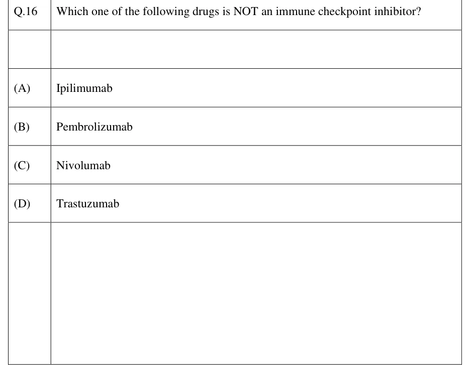
Which one of the following drugs is NOT an immune checkpoint inhibitor?
Ipilimumab
Pembrolizumab
Nivolumab
Trastuzumab
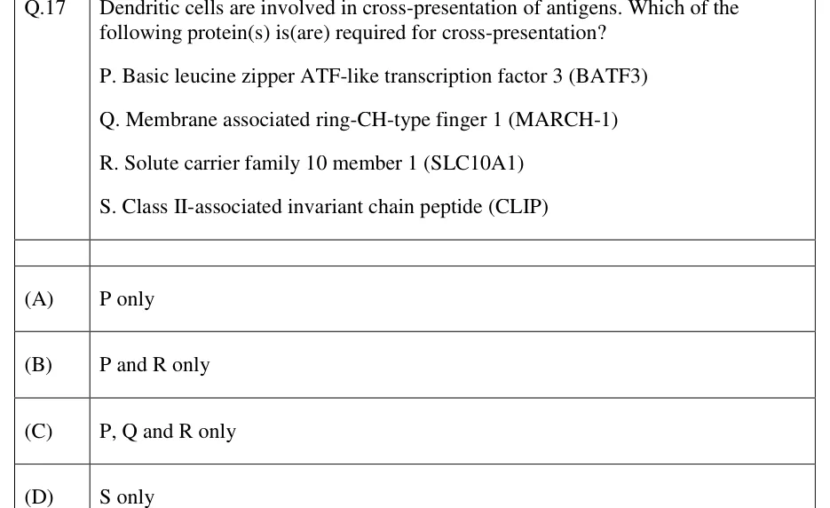
Dendritic cells are involved in cross-presentation of antigens. Which of the following protein(s) is(are) required for cross-presentation?
P. Basic leucine zipper ATF-like transcription factor 3 (BATF3)
Q. Membrane associated ring-CH-type finger 1 (MARCH-1)
R. Solute carrier family 10 member 1 (SLC10A1)
S. Class II-associated invariant chain peptide (CLIP)
P only
P and R only
P, Q and R only
S only
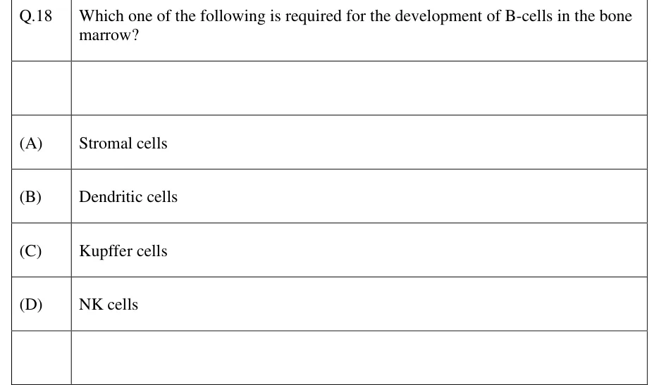
Which one of the following is required for the development of B-cells in the bone marrow?
Stromal cells
Dendritic cells
Kupffer cells
NK cells
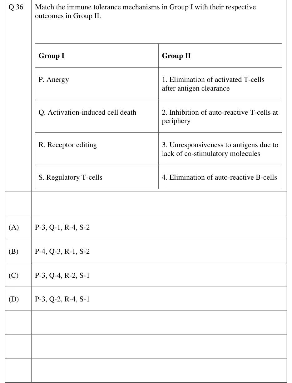
Match the immune tolerance mechanisms in Group I with their respective outcomes in Group II.
Group I: P. Anergy; Q. Activation-induced cell death; R. Receptor editing; S. Regulatory T-cells.
Group II: 1. Elimination of activated T-cells after antigen clearance; 2. Inhibition of auto-reactive T-cells at periphery; 3. Unresponsiveness to antigens due to lack of co-stimulatory molecules; 4. Elimination of auto-reactive B-cells.
P-3, Q-1, R-4, S-2
P-4, Q-3, R-1, S-2
P-3, Q-4, R-2, S-1
P-3, Q-2, R-4, S-1
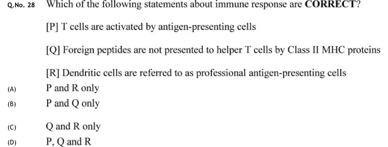
Which of the following statements about immune response are CORRECT?
[P] T cells are activated by antigen-presenting cells
[Q] Foreign peptides are not presented to helper T cells by Class II MHC proteins
[R] Dendritic cells are referred to as professional antigen-presenting cells
P and R only
P and Q only
Q and R only
P, Q and R
PRACTICE
A virus-infected cell presents a viral peptide to cytotoxic T cells. Which protein moves the peptide from the cytosol into the ER lumen for loading onto MHC class I?
TAP (transporter associated with antigen processing)
Invariant chain (Ii)
HLA-DM
Cathepsin S
Answer: (A)
Cytosolic peptides produced by the proteasome must enter the ER, where class I is loaded.
This transport is done by TAP1/TAP2.
Invariant chain and HLA-DM belong to the class II pathway, and cathepsins act in endosomes.
Which of the following statements about thymocyte selection are correct?
Positive selection occurs in the thymic cortex and requires weak binding of the TCR to self-MHC.
Negative selection removes thymocytes with high-affinity TCRs for self-peptide–MHC.
AIRE promotes expression of tissue-restricted antigens in medullary thymic epithelial cells.
Double-negative thymocytes express both CD4 and CD8.
Answer: (A), (B), (C)
A is correct: cortical epithelium gives the survival signal to low-to-moderate affinity binders, and cells that fail die by neglect.
B is correct: strongly self-reactive cells undergo apoptosis.
C is correct: AIRE drives display of peripheral self-antigens in the medulla.
D is wrong: double-negative cells lack both; double-positive cells have both.
The parents carry four different HLA haplotypes between them: mother a/b and father c/d. A patient is a/c and needs a haploidentical or better donor. Ignoring recombination, what is the probability (as a decimal) that a new full sibling shares at least one haplotype with the patient?
Answer: (accepted range )
A new sibling shares no haplotype only if it receives b from the mother and d from the father: 1/2 × 1/2 = 1/4.
Probability of sharing at least one = 1 − 1/4 = 3/4 = 0.75.
A disease has . A vaccine has efficacy 0.90 in preventing transmission. Assuming homogeneous mixing, what percentage of the population must be vaccinated to reach herd immunity? Give the answer to one decimal place.
Answer: (accepted range )
Immune fraction needed = 1 − 1/3 = 2/3 = 0.6667.
Coverage = 0.6667 / 0.90 = 0.7407.
As a percentage this is about 74.1.
Which of the following pairings of hypersensitivity type and example is correct?
Type I: anaphylaxis to peanut, mediated by IgE bound to mast cells
Type II: serum sickness, caused by immune complexes
Type III: haemolytic transfusion reaction, caused by antibody to red-cell antigens
Type IV: contact dermatitis, mediated by IgG and complement
Answer: (A)
A is correct: type I is IgE and mast-cell mediated.
B is wrong: serum sickness is type III.
C is wrong: a haemolytic transfusion reaction is type II.
D is wrong: type IV is T-cell mediated, not IgG and complement.
OFFICIAL GATE 2027 SYLLABUS WORDING
Copied from the GATE 2027 syllabus; see all 2027 syllabi.
GATE BT
T-cell and B-cell development; Memory responses; Major Histocompatibility complex (MHC); Antigen processing and presentation; Regulation of immune responses; Immune tolerance; Hypersensitivity; Autoimmunity and Immunodeficiency; Graft vs Host disease; Immunization and vaccines
Preparing in Kolkata? IAE Academy's course for GATE BT.