IAE Academy

GATE BT

Immune system

Lymphoid organs, innate immunity, complement and antibodies

The immune system is organised into primary and secondary lymphoid organs. Its fast innate arm (barriers, pattern-recognition receptors, phagocytes, NK cells, inflammation, complement) signals through cytokines and chemokines and primes the slower adaptive arm, whose B cells make antibodies of enormous diversity. GATE questions test where each cell matures or meets antigen, which receptor or pathway does what, the three complement cascades, antibody structure and classes, the DNA-level mechanisms (V(D)J recombination, somatic hypermutation, class switching), and hybridoma technology.

6 previous-year questions · 6 solved · 5 practice questions

Lymphoid organs and the cells of the immune system

All immune cells arise from haematopoietic stem cells. Primary (central) lymphoid organs are where lymphocytes are produced and become mature but antigen-naive. Secondary (peripheral) lymphoid organs are where naive lymphocytes meet antigen and where adaptive responses start.

Organ

Type

Main role

Bone marrow

Primary

Site of all blood-cell production; B-cell development and maturation (in humans and mice); NK-cell development

Thymus

Primary

T-cell maturation: positive selection in the cortex (recognise self-MHC), negative selection mainly at the cortico-medullary junction and medulla (delete strongly self-reactive cells); involutes with age

Bursa of Fabricius

Primary (birds only)

B-cell maturation in birds; mammals have no bursa

Lymph nodes

Secondary

Filter lymph; B-cell follicles (germinal centres after activation) in the cortex, T cells in the paracortex, plasma cells in the medulla; antigen arrives by afferent lymphatics, free or carried by dendritic cells

Spleen

Secondary

Filters blood (not lymph). White pulp: T cells around the central arteriole (PALS) and B-cell follicles; red pulp removes old red cells

MALT (GALT, BALT; Peyer's patches, tonsils, appendix)

Secondary

Respond to antigen at mucosal surfaces; M cells carry antigen across the epithelium of Peyer's patches

Cells of the myeloid lineage (neutrophils, eosinophils, basophils, mast cells, monocytes/macrophages, most dendritic cells) mainly drive innate responses. Cells of the lymphoid lineage are B cells, T cells (CD4 helper, CD8 cytotoxic) and NK cells. Dendritic cells are the most efficient antigen-presenting cells for activating naive T cells, and they link innate and adaptive immunity.

EXAM TIP

Remember the flow of antigen: lymph nodes sample lymph, the spleen samples blood, MALT samples mucosal surfaces. The thymus and bone marrow are where lymphocytes develop. Adaptive responses to a foreign antigen normally start in the secondary organs, although the bone marrow later houses long-lived plasma cells.

Innate immunity and inflammation

Innate immunity is immediate, uses germline-encoded receptors and has no antigen-specific memory. The first lines are barriers: skin, mucus, cilia, low gastric pH, lysozyme, defensins and commensal flora. When these are breached, pattern-recognition receptors (PRRs) detect conserved microbial molecules (PAMPs) or damage signals (DAMPs).

Receptor

Location

Ligand

TLR4 (with MD-2, CD14)

Cell surface

Lipopolysaccharide (LPS) of Gram-negative bacteria

TLR2 (with TLR1 or TLR6)

Cell surface

Lipoproteins, peptidoglycan, lipoteichoic acid

TLR5

Cell surface

Flagellin

TLR3

Endosome

Double-stranded RNA

TLR7 and TLR8

Endosome

Single-stranded RNA

TLR9

Endosome

Unmethylated CpG DNA

RIG-I-like receptors

Cytosol

Viral RNA

NOD1 and NOD2 (NOD-like receptors)

Cytosol

Fragments of bacterial peptidoglycan

Inflammasome-forming NLRs (for example NLRP3)

Cytosol

Microbial products and danger signals; the inflammasome activates caspase-1, which processes pro-IL-1β and pro-IL-18 into active cytokines

TLR signalling through adaptor proteins activates NF-κB, which switches on genes for inflammatory cytokines (TNF-α, IL-1, IL-6) and co-stimulatory molecules. All TLRs except TLR3 use the adaptor MyD88; TLR3 uses TRIF, and TLR4 can use both. Sensing of viral nucleic acid also activates IRF transcription factors and type I interferons.

Phagocytosis, in order

  1. Recognition of the microbe, directly by PRRs or through opsonins (antibody, C3b)

  2. Engulfment into a phagosome

  3. Fusion with a lysosome to form a phagolysosome

  4. Killing by the respiratory burst (NADPH oxidase makes superoxide), nitric oxide (iNOS), lysozyme and proteases

The main phagocytes are neutrophils (short-lived, first to arrive at a site of infection) and macrophages (long-lived, and they also present antigen to T cells).

NK cells are lymphocytes without antigen-specific receptors. They kill virus-infected and tumour cells that have lost MHC class I (the 'missing self' idea), because their inhibitory receptors then go unengaged, and they are triggered by activating receptors (for example NKG2D) that bind stress-induced ligands. They kill with perforin and granzymes, secrete IFN-γ, and can perform antibody-dependent cellular cytotoxicity (ADCC) through the Fc receptor CD16 (FcγRIII).

Inflammation shows redness, heat, swelling and pain. Mast cells and macrophages release histamine, prostaglandins, leukotrienes and cytokines; bradykinin comes from the kinin system. Vasodilation and increased vascular permeability bring fluid, complement and antibodies into the tissue. In the liver, IL-6 (with IL-1 and TNF-α) induces acute-phase proteins such as C-reactive protein, serum amyloid A, fibrinogen and mannose-binding lectin.

Neutrophil extravasation, in order

  1. Rolling on the endothelium: endothelial E- and P-selectins bind sialyl-Lewis X carbohydrates on the neutrophil

  2. Activation by chemokines bound to the endothelium

  3. Firm adhesion: integrins (LFA-1, Mac-1) bind ICAM-1

  4. Diapedesis between endothelial cells (PECAM-1 is involved)

  5. Chemotaxis up a gradient of chemokines, C5a and bacterial peptides to the site

EXAM TIP

Intracellular TLRs (3, 7, 8, 9) sit in endosomes and see nucleic acids; surface TLRs (1, 2, 4, 5, 6) see lipids, lipoproteins, LPS and flagellin.

Cytokines and chemokines

Cytokines are small secreted proteins that act on cells through specific receptors, locally (autocrine, paracrine) or sometimes systemically (endocrine). They are pleiotropic (one cytokine, many effects) and redundant (different cytokines, same effect), and they can act in synergy or antagonism. Chemokines are a family of small cytokines that direct cell migration by binding G-protein-coupled receptors; they are grouped by their conserved cysteines into CC, CXC, CX3C and C classes.

Cytokine

Main source

Key actions

IL-1, TNF-α, IL-6

Macrophages, dendritic cells

Pro-inflammatory; fever; acute-phase response (IL-6 in liver)

IL-2

Activated T cells

T-cell growth and proliferation; supports Treg and NK cells

IL-4

Th2 cells, mast cells

Drives Th2 differentiation; switching to IgE (and IgG4 in humans, IgG1 in mouse)

IL-5

Th2 cells

Eosinophil growth and activation; promotes IgA production in mice

IL-7

Bone marrow and thymic stromal cells

Development and survival of B and T lymphocytes

IL-10

Treg, macrophages, Th2

Anti-inflammatory; suppresses macrophage and Th1 function

IL-12

Macrophages, dendritic cells

Drives Th1 differentiation; makes NK cells produce IFN-γ

IL-17

Th17 cells

Induces chemokines that recruit neutrophils; defence against extracellular bacteria and fungi

IFN-γ

Th1, CD8 T cells, NK cells

Activates macrophages; raises MHC class I and class II expression; favours IgG switching

IFN-α and IFN-β (type I)

Virus-infected cells, plasmacytoid dendritic cells

Antiviral state; raise MHC class I; activate NK cells

TGF-β

Treg, many cells

Immunosuppressive; drives IgA switching; with IL-6 favours Th17

CXCL8 (IL-8)

Macrophages, endothelium

Chemokine that recruits neutrophils

CCL2

Macrophages, endothelium

Chemokine that recruits monocytes

EXAM TIP

CCR5 and CXCR4 are chemokine receptors that HIV uses as co-receptors with CD4. The IL-2 receptor α chain is CD25, a marker of activated T cells and Treg.

The complement system

Complement is a set of plasma proteins, made mainly by the liver, that are activated in a cascade of proteolytic cleavages. Convention: the smaller fragment of a cleaved component is 'a' and the larger is 'b' (so C3a is small and C3b large). The exception here is C2a, which is taken as the larger fragment, giving the classical convertase as C4b2a. Some texts instead call the larger C2 fragment C2b and write the convertase as C4b2b. All three pathways converge on cleavage of C3.

Feature

Classical

Lectin

Alternative

Trigger

Antigen-bound IgM or IgG (C1q binds the Fc region); also CRP

Mannose-binding lectin or ficolins on microbial carbohydrates

Spontaneous slow hydrolysis of C3 ('tickover'); microbial surfaces lacking regulators

Early proteins

C1q, C1r, C1s, C4, C2

MBL/ficolin, MASP-1, MASP-2, C4, C2

C3, factor B, factor D, properdin

C3 convertase

C4b2a

C4b2a

C3bBb (stabilised by properdin)

C5 convertase

C4b2a3b

C4b2a3b

C3bBb3b

Needs antibody?

Yes (mainly)

No

No

C1q is activated only when it binds at least two Fc regions close together. One antigen-bound pentameric IgM is therefore enough, but IgG must be bound at two neighbouring sites on the antigen.

C3 CONVERTASES

Each cleaves C3 into C3a (anaphylatoxin) and C3b (opsonin); C3b joining the convertase makes the C5 convertase.

TERMINAL PATHWAY AND MEMBRANE ATTACK COMPLEX

Common to all pathways. The MAC is a pore that lyses Gram-negative bacteria and other susceptible cells.

Effector function

Mediated by

Opsonisation

C3b (and C4b) bound to the microbe, recognised by complement receptor CR1 on phagocytes; iC3b, recognised by CR3

Inflammation and recruitment

Anaphylatoxins C3a, C4a, C5a (C5a is the most potent); C5a is also a strong chemoattractant for neutrophils

Lysis

Membrane attack complex (C5b-C9)

Clearance of immune complexes

C3b binding, with transport by CR1 on red cells

Regulator

Action

C1 inhibitor

Blocks C1r, C1s and MASPs; deficiency causes hereditary angioedema

DAF (CD55)

Accelerates decay of C3 convertases on host cells

Factor H and factor I

Factor I cleaves C3b with cofactors (factor H, MCP/CD46); factor H also displaces Bb from the alternative convertase

C4b-binding protein

Cofactor for factor I cleavage of C4b; accelerates decay of C4b2a

CD59 (protectin)

Prevents C9 polymerisation, so blocks the MAC on host cells. CD55 and CD59 are attached by a GPI anchor; their absence on red cells (a defect in GPI-anchor synthesis) underlies paroxysmal nocturnal haemoglobinuria

EXAM TIP

Deficiency patterns: C3 deficiency gives recurrent severe pyogenic infections; deficiency of the terminal components C5-C9 gives recurrent Neisseria infections; deficiency of the early classical components (C1, C2, C4) is linked to immune-complex disease such as lupus.

Antibody structure and classes

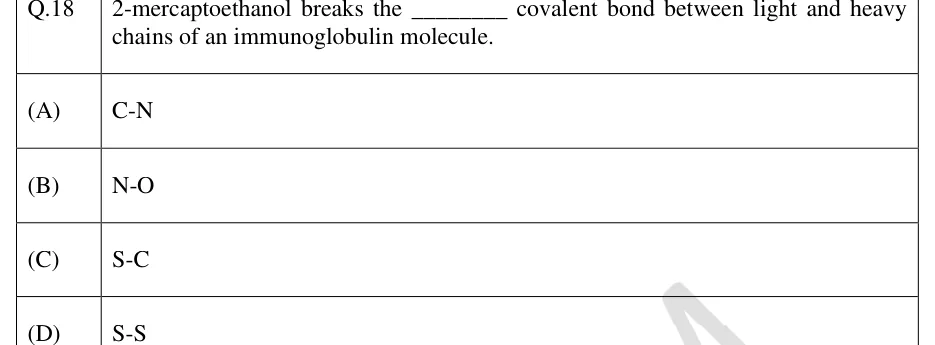
An antibody (immunoglobulin) is a Y-shaped molecule of two identical heavy (H) chains and two identical light (L) chains joined by disulphide bonds. Each chain has a variable (V) domain and constant (C) domains. The V domains of an H and an L chain together form one antigen-binding site. Within each V domain, three complementarity-determining regions (CDRs) are the hypervariable loops that contact the antigen, so each site has six CDRs. IgG, IgA and IgD have three CH domains and a flexible hinge between CH1 and CH2; IgM and IgE have four CH domains and no hinge. Light chains are κ or λ (never both in one antibody); the heavy-chain type defines the class.

APPROXIMATE SIZE OF IGG

Heavy chain about 50 kDa, light chain about 25 kDa; IgG is a monomer with 2 antigen-binding sites.

Fragments from enzymatic digestion of IgG

  1. Papain cuts above the interchain disulphides of the hinge: two Fab fragments (each with one antigen-binding site) plus one Fc fragment

  2. Pepsin cuts below the hinge disulphides: one F(ab')2 fragment (two binding sites, so it can still cross-link antigen), while the Fc is digested into small pieces

  3. The Fc region determines the biological function (complement binding, Fc-receptor binding, placental transfer)

Class (heavy chain)

Form

Key features and functions

IgG (γ)

Monomer

Most abundant in serum; main antibody of secondary responses; opsonisation, complement activation, ADCC; the only class that crosses the human placenta (via FcRn); neutralises toxins and viruses

IgM (μ)

Pentamer with J chain (10 binding sites)

First antibody made in a primary response; best complement activator; monomeric IgM is the membrane B-cell receptor

IgA (α)

Mostly monomer in serum; dimer with J chain and secretory component in secretions (4 binding sites)

Mucosal immunity: tears, saliva, milk, gut and airway secretions; neutralises without causing inflammation; made in the largest total amount per day of any class

IgE (ε)

Monomer

Binds FcεRI on mast cells and basophils; immediate hypersensitivity (allergy); defence against helminths; lowest serum level

IgD (δ)

Monomer

Co-expressed with IgM as the B-cell receptor on mature naive B cells; little known secreted function

VALENCY OF AN ANTIBODY

Applies when each monomer has two sites: IgG, IgE, IgD = 2; secretory IgA dimer = 4; IgM pentamer = 10.

Antigenic determinants on antibodies

  1. Isotype: constant-region differences that define the classes and subclasses; all normal members of a species carry every isotype

  2. Allotype: allelic variants of constant regions that differ between individuals of the same species

  3. Idiotype: the unique determinants in the variable region (mainly the CDRs) of one antibody or one B-cell clone

EXAM TIP

The J chain joins IgM pentamers and IgA dimers. The secretory component is added when IgA crosses the epithelium and comes from the poly-Ig receptor. The membrane form of each class has a hydrophobic transmembrane tail and the secreted form ends in a hydrophilic tail; the two arise by alternative RNA processing.

Molecular basis of antibody diversity

The body makes an enormous repertoire of antibodies from a limited number of genes. In the germline, the genes for the V region are split into segments. In developing B cells, V(D)J recombination joins one V, one D and one J segment for the heavy chain, and one V and one J segment for each light chain (κ or λ). Light chains have no D segments.

Features of V(D)J recombination

  1. Recombination signal sequences (a conserved heptamer and nonamer separated by a 12-bp or 23-bp spacer) flank each segment

  2. 12/23 rule: a segment with a 12-bp spacer joins only a segment with a 23-bp spacer

  3. RAG-1 and RAG-2 make the double-strand breaks (lymphocyte-specific; also used in T-cell receptor rearrangement); Artemis opens the hairpin ends and non-homologous end joining repairs the break

  4. Junctional diversity: exonuclease trimming, P nucleotides (palindromic, from hairpin opening) and N nucleotides added without a template by terminal deoxynucleotidyl transferase (TdT). N nucleotides are added mainly at heavy-chain junctions, because TdT is expressed while the heavy chain rearranges

  5. Allelic exclusion: only one heavy-chain allele and one light-chain allele is productively expressed per B cell, and only one light-chain type (κ or λ), so each B cell makes one specificity

  6. Order: the heavy chain rearranges first (D to J, then V to DJ), then the light chain, κ before λ

COMBINATORIAL DIVERSITY

Counts of functional segments. Total combinations are N_H x N_L (assuming any heavy chain pairs with any light chain), before junctional diversity and somatic hypermutation are added.

Sources of antibody diversity

  1. Multiple germline V, D and J segments

  2. Combinatorial joining of V(D)J segments

  3. Junctional diversity (trimming, P and N nucleotides)

  4. Combinatorial pairing of heavy and light chains

  5. Somatic hypermutation after antigen stimulation

After antigen activates a mature B cell (with T-cell help) in a germinal centre, two further changes occur. Both need activation-induced cytidine deaminase (AID), which deaminates cytosine to uracil in DNA; RAG proteins are not involved.

Feature

Somatic hypermutation

Class (isotype) switching

What changes

Point mutations in the rearranged V(D)J exons of H and L chains

The heavy-chain constant region (class) changes

Effect on antigen specificity

Alters affinity (up or down)

None: the same VDJ is joined to a different C gene

DNA event

Random point mutations at about per base pair per cell division, roughly a million times the background rate

Switch regions upstream of the C genes recombine; the DNA between them is looped out and deleted

Needs

AID, T-cell help, germinal centre

AID, CD40-CD40L interaction, cytokines

Outcome

Selection of B cells with higher-affinity receptors raises affinity over time (affinity maturation)

Changes effector function; one-way (irreversible, since DNA is deleted)

Cytokines direct which class is chosen: IL-4 leads to IgE (and IgG4 in humans), IFN-γ favours IgG classes, TGF-β favours IgA. A naive B cell expresses IgM and IgD together from one long transcript by alternative splicing; this needs no DNA change. Defects in CD40L or in AID cause hyper-IgM syndrome, with high IgM and little IgG, IgA or IgE.

WORKED EXAMPLE

A hypothetical B-cell heavy-chain locus has 40 functional V, 25 D and 6 J segments. The κ locus has 35 V and 5 J segments, and the λ locus has 30 V and 4 J segments. Assuming any heavy chain can pair with any light chain, how many different antibodies can form by combinatorial joining alone?

Solution
  1. Heavy chain combinations: 40 x 25 x 6 = 6000

  2. Light chain combinations: κ 35 x 5 = 175; λ 30 x 4 = 120; total 175 + 120 = 295

  3. Any heavy chain can pair with any light chain: 6000 x 295 = 1 770 000

Answer: 1 770 000 combinations (junctional diversity and somatic hypermutation would add to this)

EXAM TIP

RAG gives V(D)J joining in developing B cells (bone marrow). AID gives hypermutation and switching in mature, activated B cells (germinal centre). Do not swap them.

Effector responses and polyclonal and monoclonal antibodies

Humoral immunity is mediated by antibodies: neutralisation of toxins and viruses, opsonisation for phagocytosis, complement activation and ADCC. Most protein antigens need T-cell help: a CD4 follicular helper T cell recognises peptide on the B cell's MHC class II and delivers CD40L and cytokines (such as IL-21). This leads to antibody production, memory B cells and plasma cells. Cell-mediated immunity is mediated by T cells and the macrophages they activate.

MHC class I is on almost all nucleated cells and presents peptides from cytosolic (endogenous) proteins to CD8 T cells. MHC class II is on professional antigen-presenting cells (dendritic cells, macrophages, B cells) and presents peptides from endocytosed (exogenous) proteins to CD4 T cells.

T cell

Driving cytokines

Master transcription factor

Signature cytokines

Main role

Th1

IL-12, IFN-γ

T-bet

IFN-γ

Activate macrophages; defence against intracellular microbes

Th2

IL-4

GATA-3

IL-4, IL-5, IL-13

Defence against helminths; IgE; allergy

Th17

TGF-β, IL-6 (IL-23 sustains them)

RORγt

IL-17, IL-22

Neutrophil recruitment; extracellular bacteria and fungi

Treg

TGF-β, IL-2

FoxP3

IL-10, TGF-β

Suppress responses; tolerance

Cytotoxic CD8 (CTL)

IL-2 (from CD4 help), IL-12

T-bet, Eomesodermin

IFN-γ, TNF-α

Recognise peptide on MHC class I; kill by perforin and granzymes, and by Fas-FasL

Feature

Primary antibody response

Secondary antibody response

Responding B cells

Naive B cells

Memory B cells

Lag before antibody appears

Longer

Shorter

Peak antibody level

Lower

Higher

Main class

IgM first, then IgG

Mainly IgG (also IgA, IgE)

Average affinity

Lower

Higher (affinity maturation)

Antigen needed

Higher dose; adjuvants often needed for proteins

Lower dose

T-independent antigens, such as bacterial polysaccharides and LPS, carry many repeated epitopes that cross-link B-cell receptors and activate B cells without T-cell help. The response is mainly IgM, with little class switching, little affinity maturation and little memory.

A polyclonal antibody preparation (for example, antiserum from an immunised animal) is a mixture of antibodies from many B-cell clones, recognising many epitopes of one antigen. A monoclonal antibody comes from a single B-cell clone: every molecule is identical and binds one epitope.

Feature

Polyclonal

Monoclonal

Source

Serum of an immunised animal

One B-cell clone (for example a hybridoma)

Epitopes recognised

Many epitopes of the antigen

One epitope

Batch-to-batch consistency

Varies between animals and bleeds

Identical; unlimited supply from the clone

Tolerance of small changes in the antigen

Higher (other epitopes still bind)

Lower (binding lost if the one epitope changes)

Cost and time to produce

Lower, faster

Higher, slower

Hybridoma production of monoclonal antibodies

  1. Immunise a mouse with the antigen; collect spleen B cells

  2. Fuse them (for example with polyethylene glycol) with myeloma cells that are immortal but lack the enzyme HGPRT and do not secrete their own antibody

  3. Grow in HAT medium (hypoxanthine, aminopterin, thymidine): aminopterin inhibits dihydrofolate reductase and so blocks de novo synthesis of purines and thymidylate. Cells must then use the salvage pathways: HGPRT for hypoxanthine and thymidine kinase for thymidine

  4. Unfused myeloma cells die (no HGPRT); unfused B cells die (they do not survive long in culture); only hybrids (HGPRT from the B cell, immortality from the myeloma) grow

  5. Screen supernatants (for example by ELISA) and clone by limiting dilution to obtain one clone

Monoclonal antibodies are used in diagnostics (ELISA, immunofluorescence, flow cytometry, pregnancy tests), in affinity purification and in therapy. Mouse antibodies trigger an immune response in humans, so therapeutic antibodies are engineered: chimeric (mouse V region, human C region), humanised (only the CDRs, with a few framework residues, from mouse) or fully human (from transgenic mice or phage display). The naming scheme used for most antibodies approved so far marks these with the endings -omab (mouse), -ximab (chimeric), -zumab (humanised) and -umab (human); newer names follow a revised scheme without these markers. Polyclonal antibodies are used for antivenoms and antitoxins, and as secondary antibodies in detection.

EXAM TIP

HAT selection is a classic trap: the myeloma partner must be HGPRT-deficient. The hybrid survives because it gets HGPRT from the B cell and unlimited growth from the myeloma.

PREVIOUS-YEAR QUESTIONS

6 questions from the BT papers

Try each question first, then open the answer. We placed each question under a topic ourselves; the exam paper does not label topics.

See these papers in full: GATE BT 2026 · GATE BT 2024 · GATE BT 2022 · GATE BT 2020 · GATE BT 2019

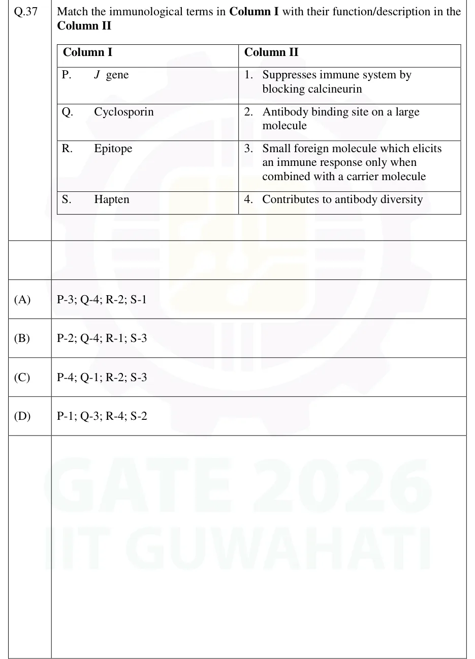
GATE 2026 BT Q.37

GATE 2026 · BTQ.37 · 2 marks · MCQ

Match the immunological terms in Column I with their function/description in the Column II. Column I: P. J gene; Q. Cyclosporin; R. Epitope; S. Hapten. Column II: 1. Suppresses immune system by blocking calcineurin; 2. Antibody binding site on a large molecule; 3. Small foreign molecule which elicits an immune response only when combined with a carrier molecule; 4. Contributes to antibody diversity.

  1. (A)

    P-3; Q-4; R-2; S-1

  2. (B)

    P-2; Q-4; R-1; S-3

  3. (C)

    P-4; Q-1; R-2; S-3

  4. (D)

    P-1; Q-3; R-4; S-2

Answer and solution
See the question in the paper
GATE 2026 BT Q.37: answer and solution

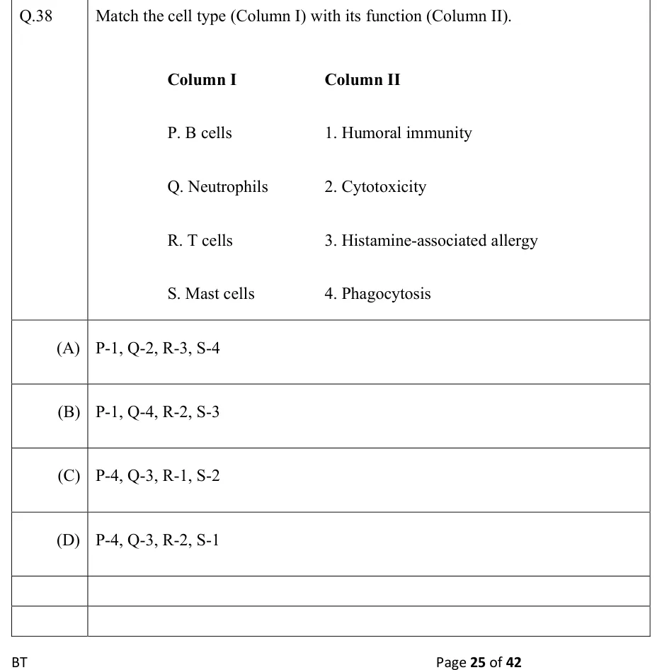
GATE 2022 BT Q.38

GATE 2022 · BTQ.38 · 2 marks · MCQ

Match the cell type (Column I) with its function (Column II).

Column I: P. B cells; Q. Neutrophils; R. T cells; S. Mast cells
Column II: 1. Humoral immunity; 2. Cytotoxicity; 3. Histamine-associated allergy; 4. Phagocytosis

  1. (A)

    P-1, Q-2, R-3, S-4

  2. (B)

    P-1, Q-4, R-2, S-3

  3. (C)

    P-4, Q-3, R-1, S-2

  4. (D)

    P-4, Q-3, R-2, S-1

Answer and solution
See the question in the paper
GATE 2022 BT Q.38: answer and solution

PRACTICE

5 questions to test yourself

PRACTICE 11 mark · MCQ

At which complement component do the classical, lectin and alternative pathways converge, with its larger cleavage fragment acting as an opsonin?

  1. (A)

    C1q

  2. (B)

    C3

  3. (C)

    C5

  4. (D)

    C9

Answer and solution

Answer: (B)

  1. All three pathways form a C3 convertase (C4b2a or C3bBb), so they converge on cleavage of C3.

  2. C3 is split into C3a (anaphylatoxin) and C3b (opsonin).

  3. C1q is classical only, C5 is cleaved later by the C5 convertase, and C9 polymerises in the MAC.

PRACTICE 22 marks · MCQ

In hybridoma production, spleen cells from an immunised mouse are fused with myeloma cells lacking HGPRT and grown in HAT medium. Which cells survive?

  1. (A)

    Unfused myeloma cells only

  2. (B)

    Unfused spleen B cells only

  3. (C)

    Fused spleen cell-myeloma hybrids only

  4. (D)

    All cells, because hypoxanthine rescues the myeloma cells

Answer and solution

Answer: (C)

  1. Aminopterin blocks de novo nucleotide synthesis, so cells must use the salvage pathway.

  2. Unfused myeloma cells lack HGPRT, so they cannot salvage hypoxanthine and die.

  3. Unfused B cells have HGPRT but do not survive long in culture.

  4. Hybrids have HGPRT from the B cell and immortality from the myeloma, so they survive.

PRACTICE 32 marks · MSQ

Which of the following statements about immunoglobulin class switching are correct?

  1. (A)

    It requires activation-induced cytidine deaminase (AID).

  2. (B)

    It changes the antigen specificity of the antibody.

  3. (C)

    The DNA between the switch regions is deleted.

  4. (D)

    It requires RAG-1 and RAG-2.

Answer and solution

Answer: (A), (C)

  1. Class switching uses AID at switch regions, and the intervening DNA is looped out and lost, so it is irreversible (A and C true).

  2. The same VDJ exon is kept, so antigen specificity is unchanged (B false).

  3. RAG proteins act in V(D)J recombination in developing B cells, not in switching (D false).

PRACTICE 42 marks · NAT

A heavy-chain locus has 50 functional V, 20 D and 5 J segments. The κ locus has 40 V and 5 J segments and the λ locus has 30 V and 4 J segments. Considering only combinatorial joining of segments, and assuming every heavy chain can pair with every light chain and each pair is a distinct specificity, how many different antibody specificities can be formed?

Answer and solution

Answer:

  1. Heavy chain: 50 x 20 x 5 = 5000.

  2. Light chain: 40 x 5 + 30 x 4 = 200 + 120 = 320.

  3. Total: 5000 x 320 = 1 600 000.

PRACTICE 51 mark · NAT

How many antigen-binding sites are present on one molecule of secretory IgA that is a dimer of two IgA monomers?

Answer and solution

Answer:

  1. Each IgA monomer has two antigen-binding sites.

  2. A dimer has two monomers, so 2 x 2 = 4 sites.

  3. The J chain and secretory component add no binding sites.

OFFICIAL GATE 2027 SYLLABUS WORDING

Copied from the GATE 2027 syllabus; see all 2027 syllabi.

GATE BT

Immunology: Primary and Secondary lymphoid organs; Innate immunity and Inflammation; Cytokines and Chemokines; Complement system; Effector responses: Cellular and Humoral Immunity; Molecular basis of Antibody diversity and function; Polyclonal and Monoclonal antibodies

Preparing in Kolkata? IAE Academy's course for GATE BT.