IAE Academy

GATE BT

Biomolecules and biological membranes

This topic covers the structure and function of the four classes of biomolecules (amino acids and proteins, carbohydrates, lipids, nucleic acids) and how biological membranes control what crosses them. It matters for GATE BT because questions ask you to compute pI and charge, identify linkages and reducing sugars, apply the Nernst and Goldman equations and the free energy of transport, and reason about pumps, channels, the action potential and motor proteins.

12 previous-year questions · 11 solved · 6 practice questions

Amino acids and the peptide bond

Every standard amino acid has a central alpha carbon bonded to an amino group, a carboxyl group, a hydrogen atom and a side chain (R). Proteins are built from 20 standard amino acids. Every one except glycine has a chiral alpha carbon, and in proteins it is always the L configuration; glycine (R = H) is achiral. Proline's side chain loops back and bonds to its own alpha-amino nitrogen, making it a secondary amine (traditionally called an imino acid); its ring fixes the backbone angle , so proline often puts a kink or bend in a chain. Beyond the standard 20, a few proteins contain selenocysteine (encoded by a recoded UGA) and, in some archaea and bacteria, pyrrolysine (a recoded UAG).

Class

Amino acids (side chain at pH 7)

Nonpolar aliphatic

Gly, Ala, Val, Leu, Ile, Met, Pro

Aromatic

Phe, Tyr, Trp (Tyr is also polar; Trp and Tyr account for most of a protein's absorbance at 280 nm)

Polar, uncharged

Ser, Thr, Cys, Asn, Gln

Positively charged (basic)

Lys, Arg, His (His side-chain pKa is about 6, so only a minority of His side chains is protonated at pH 7)

Negatively charged (acidic)

Asp, Glu

In solution near neutral pH an amino acid exists as a zwitterion: the alpha-carboxyl group is and the alpha-amino group is . For an amino acid without an ionisable side chain the net charge is then zero; at pH 7 Asp and Glu carry about -1 and Lys and Arg about +1. Typical pKa values are about 2 for the alpha-carboxyl and 9 to 10 for the alpha-amino group. Side chains with ionisable groups (Asp, Glu, His, Cys, Tyr, Lys, Arg) add a third pKa.

HENDERSON-HASSELBALCH EQUATION

Ratio of deprotonated (A-) to protonated (HA) form of one ionisable group, with pKa = -log10 Ka. At pH = pKa the two forms are equal, and a group buffers best within about 1 pH unit of its pKa.

ISOELECTRIC POINT, AMINO ACID WITH A NON-IONISABLE SIDE CHAIN

pK1 is the alpha-COOH and pK2 the alpha-NH3+. For any amino acid, pI is the average of the two pKa values that lie on either side of the neutral (net charge zero) form.

Procedure to find pI of any amino acid

  1. Write the fully protonated form and its net charge (usually +1, or +2 for Lys, Arg, His).

  2. Deprotonate groups in order of increasing pKa, updating the net charge after each.

  3. Find the species with net charge 0.

  4. Average the pKa that forms it (the one before) and the pKa that removes its next proton (the one after).

  5. Acidic amino acids (Asp, Glu): pI is the mean of the two lowest pKa values. Basic ones (Lys, Arg, His): the mean of the two highest.

  6. Cys and Tyr side chains are neutral when protonated and negative when deprotonated, so the same 'two lowest pKa values' rule applies to them.

WORKED EXAMPLE

Histidine has pKa values 1.8 (alpha-COOH), 6.0 (imidazole) and 9.2 (alpha-NH3+). Find its pI and its net charge at pH 5.0.

Solution
  1. Fully protonated form at very low pH: carboxyl neutral (0), imidazole +1, amino +1, net +2.

  2. After pKa 1.8: net +1. After pKa 6.0: net 0. After pKa 9.2: net -1.

  3. The neutral species lies between pKa 6.0 and 9.2, so pI = (6.0 + 9.2)/2 = 7.6.

  4. At pH 5.0 the carboxyl is essentially fully ionised (pH is far above 1.8) and the amino group fully protonated (pH is far below 9.2), so only the imidazole is partly charged; pH 5.0 is 1.0 below its pKa.

  5. For the imidazole, [HA+]/[A] = 10^(pKa - pH) = 10^(6.0 - 5.0) = 10, so the fraction protonated is 10/11 = 0.909.

  6. Net charge = (-1 carboxylate) + (+1 amino) + 0.909 = +0.91.

Answer: pI = 7.6; net charge at pH 5.0 is about +0.91.

The peptide bond is an amide link between the alpha-carboxyl carbon of one residue and the alpha-amino nitrogen of the next, formed with the loss of water. Resonance gives the C-N bond partial double-bond character (it is shorter than a C-N single bond), so the six atoms of the peptide group (C, C, O, N, H, C) lie in one plane and rotation about the C-N bond is restricted. The trans arrangement () is strongly favoured; the cis form occurs appreciably only in X-Pro bonds (the peptide bond to proline's nitrogen). Rotation is possible about N-C (angle ) and C-C (angle ); the sterically allowed (, ) combinations are shown on a Ramachandran plot, where right-handed alpha helices and beta strands fall in separate allowed regions. Peptides are written and numbered from the N-terminus to the C-terminus.

EXAM TIP

For pI questions, first decide which two pKa values bracket the zero-charge form. Using the two alpha-group values for Asp, Glu, Lys, Arg or His is the usual mistake.

Protein structure and denaturation

Level

What it is

Main stabilising interactions

Primary

Linear amino acid sequence

Peptide (covalent) bonds

Secondary

Local regular folds: alpha helix, beta sheet, turns

Backbone hydrogen bonds (C=O of one residue to N-H of another)

Tertiary

Complete 3-D fold of one chain

Hydrophobic effect (the main driving force), hydrogen bonds, ionic bonds, van der Waals forces, disulfide bonds

Quaternary

Arrangement of two or more chains (subunits)

Same non-covalent forces; sometimes inter-chain disulfides

The right-handed alpha helix has 3.6 residues per turn, a rise of 0.15 nm per residue and a pitch of 0.54 nm. The C=O of residue i hydrogen bonds to the N-H of residue i+4, so the hydrogen bonds run nearly parallel to the helix axis, and the side chains point outward. Within a helix proline is disruptive because its nitrogen has no hydrogen to donate and its ring prevents the required angle; glycine also destabilises helices because its flexibility favours other conformations. In a beta sheet, extended strands (rise about 0.33 to 0.35 nm per residue) lie side by side and hydrogen bond between strands, with side chains pointing alternately above and below the sheet. Strands may run parallel or antiparallel; in antiparallel sheets the hydrogen bonds are more nearly linear. A beta turn reverses chain direction over four residues, with the C=O of residue i bonded to the N-H of residue i+3, and often contains Gly or Pro.

ALPHA HELIX GEOMETRY

Right-handed alpha helix: 3.6 residues per turn, 0.15 nm rise per residue, i to i+4 hydrogen bonds.

Folding is governed by the amino acid sequence: a denatured protein can often refold in vitro to its native, active state, which shows that the sequence contains the information that specifies the native fold. In cells, chaperones help many proteins fold by preventing misfolding and aggregation, but they do not supply that information. In water-soluble proteins hydrophobic side chains are mostly buried and polar ones exposed. Disulfide bonds (Cys-Cys) form in oxidising environments and are the only covalent bonds among these stabilising forces.

Denaturation is loss of secondary, tertiary and quaternary structure without breaking peptide bonds, so the primary structure is unchanged. Heat, extreme pH, urea and guanidinium chloride (which disrupt hydrogen bonds and weaken the hydrophobic effect), and detergents all denature proteins. Reducing agents such as beta-mercaptoethanol or dithiothreitol break disulfide bonds. Denaturation is usually accompanied by loss of function and decreased solubility.

Protein

Structural point

Collagen

Three left-handed polyproline II-type chains wound around each other into a right-handed triple helix; repeat Gly-X-Y with Gly at every third position (only Gly fits at the crowded centre of the triple helix), X often Pro and Y often 4-hydroxyproline, whose formation needs vitamin C (ascorbate)

Haemoglobin

Tetramer of two alpha and two beta subunits (quaternary structure); cooperative oxygen binding

Silk fibroin

Mostly antiparallel beta sheets

EXAM TIP

Acid hydrolysis or proteases break peptide bonds; urea, heat or detergents do not. If a question says the peptide bonds remain intact, it is describing denaturation.

Carbohydrates and lipids

Monosaccharides are polyhydroxy aldehydes (aldoses) or ketones (ketoses). Glucose is an aldohexose; fructose is a ketohexose. A sugar with n chiral carbons in its open-chain form has stereoisomers, provided none is a meso form. D and L refer to the configuration at the chiral carbon farthest from the carbonyl, and natural sugars are mostly D. Epimers differ at only one chiral carbon: glucose and galactose differ at C4, glucose and mannose at C2.

In solution, pentoses and hexoses cyclise: an aldose forms a hemiacetal and a ketose a hemiketal, giving six-membered pyranose or five-membered furanose rings. The former carbonyl carbon (C1 of an aldose, C2 of a ketose) becomes the anomeric carbon, and the two configurations at it, alpha and beta, are anomers. For a D-sugar drawn as a Haworth projection, alpha has the anomeric OH on the opposite side of the ring from the terminal CH2OH group (below the ring) and beta has it on the same side (above). The anomers interconvert through the open-chain form, and the resulting change in optical rotation of a freshly dissolved sample is called mutarotation; at equilibrium D-glucose is about one-third alpha and two-thirds beta. A sugar whose anomeric carbon is free (not in a glycosidic bond) can open to the carbonyl form and reduce mild oxidising agents such as alkaline Cu2+ reagents; it is a reducing sugar. Ketoses such as fructose also test as reducing, because under the alkaline test conditions they isomerise to aldoses.

STEREOISOMERS OF A SUGAR

n is the number of chiral carbons in the open-chain form (valid when there are no meso forms). Aldohexoses have n = 4, giving 16 (8 D and 8 L); ketohexoses have n = 3, giving 8. Cyclisation adds one more chiral centre, the anomeric carbon.

Carbohydrate

Units and linkage

Reducing?

Maltose

Glucose alpha(1-4) glucose

Yes

Lactose

Galactose beta(1-4) glucose

Yes

Sucrose

Glucose alpha(1-2) beta fructose (both anomeric carbons used)

No

Trehalose

Glucose alpha(1-1) alpha glucose (both anomeric carbons used)

No

Cellobiose

Glucose beta(1-4) glucose

Yes

Amylose (starch)

Glucose alpha(1-4), unbranched, coils into a helix

One reducing end per chain

Amylopectin (starch)

Alpha(1-4) chains with alpha(1-6) branch points roughly every 24 to 30 residues

One reducing end; many non-reducing ends

Glycogen

Same linkages as amylopectin, but branched about every 8 to 12 residues (more compact)

One reducing end; many non-reducing ends

Cellulose

Glucose beta(1-4), straight chains held together by inter-chain hydrogen bonds

One reducing end per chain

Chitin

N-acetylglucosamine beta(1-4)

One reducing end per chain

Starch (plants) and glycogen (animals) are storage polysaccharides; cellulose and chitin are structural. Humans make no enzyme that hydrolyses the beta(1-4) bond between two glucose units, so cellulose is not digested (lactase cleaves the beta(1-4) galactose-glucose bond of lactose but does not act on cellulose). Glycogen's dense branching gives many non-reducing ends, where enzymes add or remove glucose units, so it can be built up and mobilised rapidly.

Lipids are biological molecules that are poorly soluble in water and soluble in nonpolar solvents. Fatty acids are long hydrocarbon chains (usually an even number of carbons, 12 to 24) ending in a carboxyl group. In the notation 18:1(), 18 is the number of carbons, 1 the number of double bonds, and places the double bond between C9 and C10 counting from the carboxyl carbon (C1); omega () numbering counts from the methyl end instead. Natural unsaturated fatty acids are mostly cis. Saturated chains pack tightly; each cis double bond kinks the chain, so melting point falls as unsaturation rises (and rises with chain length). Triacylglycerols (triglycerides) are glycerol esterified with three fatty acids and store energy. Phospholipids have a polar head and two hydrophobic tails, making them amphipathic; in water they form bilayers.

Lipid

Backbone / structure

Role

Triacylglycerol

Glycerol + 3 fatty acids (ester bonds); neutral and nonpolar

Energy storage (not a bilayer lipid); hydrolysed by alkali (saponification) to glycerol and soap

Glycerophospholipid

Glycerol + 2 fatty acids + phosphate + head group (choline, ethanolamine, serine, inositol)

Main membrane lipid

Sphingolipid

Sphingosine + one fatty acid (+ phosphocholine in sphingomyelin, or sugars in glycolipids)

Membranes, myelin, cell recognition

Sterol (cholesterol)

Four fused rings, a 3-hydroxyl head and a branched hydrocarbon tail; amphipathic

Modulates membrane fluidity; precursor of steroid hormones and bile acids

EXAM TIP

Sucrose and trehalose are non-reducing because their glycosidic bond uses both anomeric carbons. Maltose, lactose and cellobiose each keep one free anomeric carbon and are reducing. A branched polysaccharide still has only one reducing end.

Nucleotides and nucleic acids

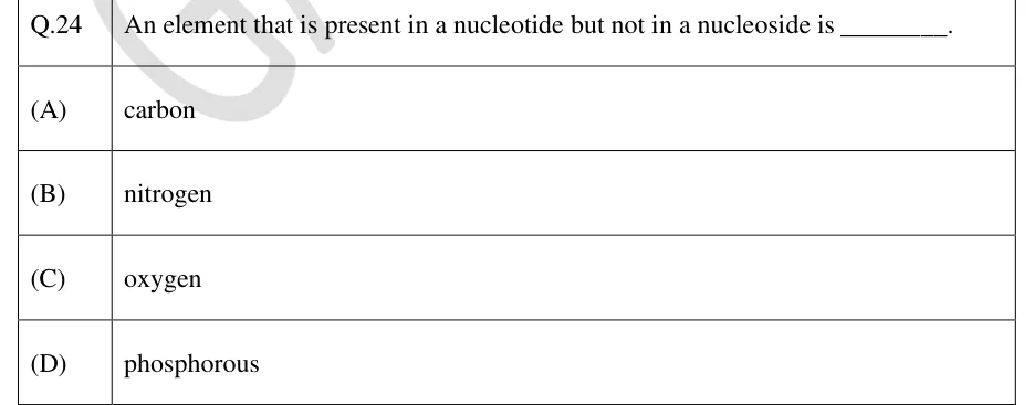
A nucleoside is a base plus a pentose sugar; a nucleotide is a nucleoside plus one or more phosphate groups (adenosine is a nucleoside; AMP, ADP and ATP are nucleotides). Purines (adenine, guanine) have two fused rings; pyrimidines (cytosine, thymine, uracil) have one. DNA uses 2'-deoxyribose and the bases A, G, C, T; RNA uses ribose and has U in place of T. The base joins the sugar's C1' by a beta-N-glycosidic bond (through N9 of a purine or N1 of a pyrimidine), and the phosphate is esterified to C5'. The 2'-OH of ribose makes RNA susceptible to alkaline hydrolysis, which DNA resists.

Nucleotides are linked by 3'-5' phosphodiester bonds, so a strand has a 5' end (usually carrying a phosphate) and a 3' end (free 3'-OH). Sequences are written 5' to 3'. In the double helix the two strands are antiparallel and complementary, with the negatively charged sugar-phosphate backbone outside. They are held together by hydrogen bonds between bases (A with T by 2 bonds, G with C by 3) and by stacking interactions between neighbouring base pairs, which contribute most of the helix's stability.

Chargaff's rules for double-stranded DNA

  1. The amount of A equals T, and G equals C.

  2. Therefore purines equal pyrimidines: A + G = T + C.

  3. The ratio (A+T)/(G+C) varies between species; within a duplex it is the same for each strand.

  4. Within one strand A need not equal T. Instead each base equals its partner in the other strand (A in one strand = T in the other), and (A+G)/(T+C) of one strand is the reciprocal of that of the other strand.

  5. These rules do not hold for single-stranded DNA or for single-stranded RNA.

B-DNA LENGTH AND TURNS

Idealised B-form: 0.34 nm rise per base pair, 10 bp per turn, so pitch 3.4 nm; diameter about 2 nm; right-handed. B-DNA in solution has about 10.5 bp per turn; use 10 unless a question states otherwise.

Form

Handedness

bp per turn

Notes

B-DNA

Right

About 10 (10.5 in solution)

Standard form in cells; wide major groove and narrow minor groove

A-DNA

Right

About 11

Dehydrated DNA; also the conformation of double-stranded RNA and RNA-DNA hybrids

Z-DNA

Left

12

Zig-zag backbone; favoured by alternating purine-pyrimidine runs

WORKED EXAMPLE

A double-stranded B-DNA fragment of 4,000 base pairs contains 28% adenine. Find the number of guanine residues, its length and the number of helical turns (take 10 bp per turn).

Solution
  1. Total nucleotides = 2 x 4,000 = 8,000.

  2. A = T = 28% each, so A + T = 56% and G + C = 44%.

  3. G = C = 22% of 8,000 = 1,760 guanines.

  4. Length = 4,000 x 0.34 nm = 1,360 nm = 1.36 micrometres.

  5. Turns = 4,000 / 10 = 400.

Answer: 1,760 guanines; length 1.36 micrometres; 400 turns.

RNA

Features

mRNA

Carries the coding sequence; eukaryotic mRNA has a 5' cap (7-methylguanosine) and a 3' poly(A) tail

tRNA

Typically 73 to 93 nucleotides (about 76); cloverleaf secondary structure, L-shaped tertiary structure; anticodon loop and a 3' CCA end to which the amino acid is attached

rRNA

Most abundant RNA; forms the core of the ribosome, and its large-subunit RNA catalyses peptide bond formation

snRNA, snoRNA

Splicing (snRNA) and rRNA modification (snoRNA)

miRNA, siRNA

Short regulatory RNAs that silence genes

EXAM TIP

The melting temperature (Tm, the temperature at which half the duplex is denatured) rises with G+C content, because G-C pairs have three hydrogen bonds and stack more strongly, and it also rises with salt concentration. Single-stranded DNA absorbs more at 260 nm than double-stranded DNA, so absorbance rises as DNA melts (hyperchromic effect).

Membrane structure and transport

The fluid mosaic model describes a membrane as a fluid phospholipid bilayer in which proteins float, and lipids and proteins diffuse laterally. Hydrophobic tails face inward and polar heads face the water. The two leaflets differ in composition (asymmetry). Lateral diffusion within a leaflet is rapid, while transverse movement of a lipid from one leaflet to the other (flip-flop) is very slow unless catalysed by flippases, floppases or scramblases. Carbohydrates, attached to lipids and proteins, lie only on the extracellular face of the plasma membrane (the side facing away from the cytosol).

Integral proteins are embedded in the bilayer and need detergents to release them; many span it with hydrophobic alpha helices of about 20 residues, and some (porins, in the outer membranes of bacteria, mitochondria and chloroplasts) form beta barrels. Peripheral proteins bind the surface by ionic and hydrogen bonds and can be released with high salt or a pH change. Fluidity increases with more unsaturated and shorter fatty acid chains and with higher temperature. Cholesterol acts as a buffer: it reduces fluidity at high temperature and prevents tight packing (keeps the membrane fluid) at low temperature.

Mechanism

Energy

Protein needed?

Direction

Example

Simple diffusion

None

No

Down gradient; not saturable

O2, CO2, small nonpolar molecules

Channel (facilitated)

None

Yes

Down electrochemical gradient; very fast; many are gated, others (leak channels, aquaporins) stay open

Aquaporin, voltage-gated K+ channel

Carrier (facilitated)

None

Yes

Down gradient; saturable (shows Jmax and Kt, like Vmax and Km); uniport

GLUT glucose transporter

Primary active

ATP hydrolysis (or light/redox)

Yes

Against gradient

Na+/K+-ATPase, SERCA Ca2+-ATPase, H+/K+-ATPase

Secondary active

Gradient of another ion

Yes

One solute uphill, driven by another moving downhill; symport (same direction) or antiport (opposite)

SGLT Na+-glucose symporter, Na+/Ca2+ exchanger

FICK'S FIRST LAW

Flux J (amount per area per time) of a solute diffusing down its concentration gradient; D is the diffusion coefficient. Across a membrane of thickness this becomes , with permeability coefficient , where K is the solute's lipid-water partition coefficient.

CARRIER-MEDIATED (FACILITATED) TRANSPORT

Same form as Michaelis-Menten kinetics: flux saturates at Jmax, and Kt is the solute concentration giving half-maximal flux. Simple diffusion has no such limit; its flux rises linearly with the concentration difference.

FREE ENERGY OF MOVING A SOLUTE FROM SIDE 1 TO SIDE 2

R = 8.314 J mol K, T in kelvin, F = 96,485 C mol, z the signed charge of the solute, and in volts; is in J mol. For an uncharged solute z = 0 and only the concentration term remains. A negative value means the movement is spontaneous (passive); a positive value means it needs an energy input.

WORKED EXAMPLE

Calculate the free energy change for moving 1 mol of Na+ into a cell at 37 degrees C (310 K) when [Na+] is 145 mM outside and 10 mM inside and the membrane potential is -70 mV (inside relative to outside).

Solution
  1. Side 1 = outside, side 2 = inside, so C2/C1 = 10/145, V and z = +1.

  2. RT = 8.314 x 310 = 2,577 J/mol; ln(10/145) = -2.674, so the concentration term = 2,577 x (-2.674) = -6,892 J/mol.

  3. Electrical term = zF = (+1)(96,485)(-0.070) = -6,754 J/mol.

  4. = -6,892 - 6,754 = -13,646 J/mol, about -13.6 kJ/mol.

Answer: About -13.6 kJ/mol. Na+ entry is strongly downhill and both terms favour it; this is the energy a Na+-coupled symporter such as SGLT uses to move glucose uphill.

OSMOTIC PRESSURE (DILUTE SOLUTIONS)

i is the number of particles each formula unit gives in solution (1 for glucose, about 2 for NaCl), C the molar concentration, R the gas constant (0.0821 L atm mol K gives in atm) and T in kelvin; iC is the osmolarity. Valid for ideal dilute solutions. Water moves by osmosis from lower to higher osmolarity, often through aquaporins; a cell swells in a hypotonic medium and shrinks in a hypertonic one (tonicity also depends on whether the solute can cross the membrane).

The Na+/K+-ATPase is a P-type ATPase: during its cycle it is transiently phosphorylated on an aspartate residue. In each cycle it pumps 3 Na+ out and 2 K+ in using 1 ATP, so it is electrogenic (net one positive charge leaves per cycle). Ouabain and digoxin inhibit it. It maintains the high intracellular K+ and low intracellular Na+ that set the resting potential and drive secondary active transport. The SERCA Ca2+ pump and the gastric H+/K+-ATPase are also P-type. Other ATP-driven pumps include V-type ATPases, which pump protons to acidify lysosomes and vesicles and form no phosphorylated intermediate, and ABC transporters (for example multidrug-resistance pumps that export drugs). F-type ATPases (the F1Fo ATP synthase) normally run the other way, making ATP as protons flow down their gradient; they can also hydrolyse ATP to pump protons.

EXAM TIP

Primary active transport uses ATP directly; secondary active transport uses an ion gradient that a primary pump built. SGLT works only because the Na+/K+-ATPase keeps intracellular Na+ low.

Membrane potential and the action potential

Unequal ion concentrations and selective permeability produce a voltage across the membrane. Approximate values for a mammalian cell (they vary with cell type) are 140 mM K+ inside and 5 mM outside, 10 mM Na+ inside and 145 mM outside, and 10 mM Cl- inside and 110 mM outside. The equilibrium potential of one ion is the voltage at which its electrical and chemical driving forces balance, given by the Nernst equation. Convention: membrane potentials are inside relative to outside.

NERNST EQUATION

One permeant ion at equilibrium; z is its signed charge (+1 for K+ and Na+, -1 for Cl-, +2 for Ca2+). 2.303RT/F = 61.5 mV at 37 degrees C (about 59 mV at 25 degrees C).

GOLDMAN (GOLDMAN-HODGKIN-KATZ) EQUATION

Steady-state potential (no net current) with several monovalent permeant ions, assuming a constant electric field across the membrane. P is each ion's permeability; only ratios matter. The Cl- terms are inverted because it is an anion. The result lies between the ions' Nernst potentials, closest to that of the most permeable ion; with only one permeant ion it reduces to the Nernst equation.

WORKED EXAMPLE

Taking K+ 5 mM out and 140 mM in, Na+ 145 mM out and 10 mM in, no Cl- permeability, PNa/PK = 0.02 and a temperature of 37 degrees C (use ), find E_K and the resting potential.

Solution
  1. E_K = 61.5 x log10(5/140) = 61.5 x (-1.447) = -89.0 mV.

  2. Take PK = 1, PNa = 0.02. Numerator = 5 + 0.02 x 145 = 7.9.

  3. Denominator = 140 + 0.02 x 10 = 140.2.

  4. Vm = 61.5 x log10(7.9/140.2) = 61.5 x (-1.249) = -76.8 mV.

Answer: E_K is about -89 mV and Vm is about -77 mV, slightly less negative than E_K because of the small Na+ leak.

The resting potential (about -70 mV in many neurons) is close to E_K but less negative, because the resting membrane is mostly permeable to K+ through leak channels, with a small Na+ leak. The action potential is an all-or-none, self-propagating voltage change in excitable cells:

Phases of the action potential

  1. Resting: Vm near -70 mV, voltage-gated channels closed.

  2. Depolarisation to threshold (about -55 mV): voltage-gated Na+ channels open, Na+ enters, and positive feedback drives the potential up towards E_Na.

  3. Peak (about +30 to +40 mV, below E_Na): Na+ channels inactivate, and the slower voltage-gated K+ channels are now open.

  4. Repolarisation: K+ leaves the cell through voltage-gated K+ channels, bringing Vm back down.

  5. Undershoot (after-hyperpolarisation): Vm dips below rest toward E_K while the K+ channels are still open; then they close and Vm returns to rest.

The spike is all-or-none: once threshold is reached its amplitude does not depend on stimulus strength, and a stronger stimulus raises the firing frequency instead. During the absolute refractory period (Na+ channels inactivated) no stimulus can trigger a second spike, which keeps the impulse travelling away from where it started. In the relative refractory period a stronger-than-normal stimulus is needed. Conduction velocity rises with axon diameter and with myelination: myelin insulates the axon so the impulse jumps between nodes of Ranvier (saltatory conduction), which is faster and uses less energy than continuous conduction. Tetrodotoxin blocks voltage-gated Na+ channels; tetraethylammonium blocks voltage-gated K+ channels. Channels can be voltage-gated, ligand-gated or mechanically gated.

EXAM TIP

The Na+/K+-ATPase does not generate the spike; the spike comes from passive ion flow through channels. The pump restores the gradients over time, and a few spikes barely change them.

Molecular motors

Motor proteins convert the chemical energy of ATP hydrolysis into directed movement along cytoskeletal filaments. Filaments have polarity, with a plus end and a minus end (for actin, the barbed and pointed ends), and each motor moves in one direction along its track.

Motor

Track

Direction

Function

Myosin II

Actin filament

Plus end (barbed end)

Muscle contraction (thick filaments), cytokinesis (contractile ring)

Myosin V

Actin

Plus end

Carries vesicles and organelles

Myosin VI

Actin

Minus end (exception among myosins)

Endocytosis

Kinesin (conventional, kinesin-1)

Microtubule

Plus end (usually away from the centrosome; towards the axon terminal, anterograde)

Vesicle and organelle transport; spindle function

Cytoplasmic dynein

Microtubule

Minus end (towards the centrosome; towards the cell body in axons, retrograde)

Retrograde transport, spindle positioning; works with dynactin

Axonemal dynein

Microtubule doublets

Minus end

Slides doublets past each other to bend cilia and flagella

Most kinesins move to the plus end; the kinesin-14 family is an exception and moves to the minus end. The cross-bridge cycle of myosin has four steps. ATP binds and the myosin head detaches from actin. ATP is hydrolysed (ADP and Pi stay bound), which cocks the head into a high-energy state. The head binds actin and releases Pi, triggering the power stroke. ADP is released, leaving the rigor state until a new ATP arrives. Kinesin is a dimer that walks hand over hand along a microtubule in steps of about 8 nm, hydrolysing about one ATP per step; myosin V walks along actin in steps of about 36 nm. In skeletal muscle, rising Ca2+ binds troponin (troponin C), which shifts tropomyosin off the myosin-binding sites on actin; thick and thin filaments then slide past each other (sliding filament model), shortening the sarcomere without shortening the filaments: the A band keeps its length while the I band and H zone shorten.

Rotary motors are also molecular motors: the F1Fo ATP synthase rotates as protons flow down their gradient, and the bacterial flagellar motor is driven by a proton (in some species Na+) gradient rather than by ATP.

EXAM TIP

ATP binding, not hydrolysis, detaches myosin from actin. Without ATP muscle stays locked in rigor, as in rigor mortis.

PREVIOUS-YEAR QUESTIONS

12 questions from the BT papers

Try each question first, then open the answer. We placed each question under a topic ourselves; the exam paper does not label topics.

See these papers in full: GATE BT 2026 · GATE BT 2025 · GATE BT 2024 · GATE BT 2023 · GATE BT 2022 · GATE BT 2020 · GATE BT 2019

GATE 2026 BT Q.23

GATE 2026 · BTQ.23 · 1 mark · MCQ

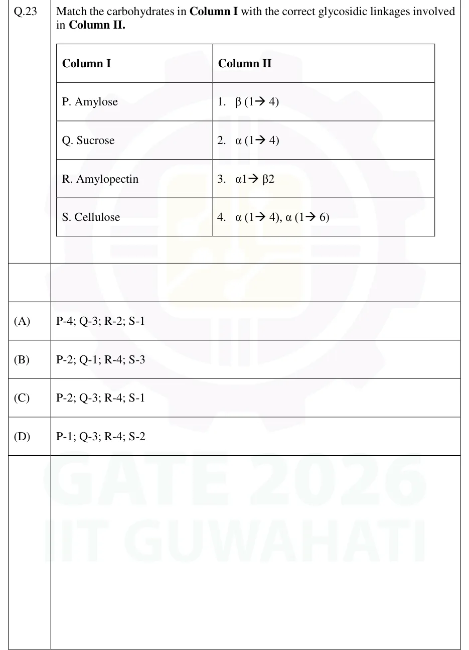
Match the carbohydrates in Column I with the correct glycosidic linkages involved in Column II.

Column I: P. Amylose; Q. Sucrose; R. Amylopectin; S. Cellulose.
Column II: 1. β (1→4); 2. α (1→4); 3. α1→β2; 4. α (1→4), α (1→6).

  1. (A)

    P-4; Q-3; R-2; S-1

  2. (B)

    P-2; Q-1; R-4; S-3

  3. (C)

    P-2; Q-3; R-4; S-1

  4. (D)

    P-1; Q-3; R-4; S-2

Answer and solution
See the question in the paper
GATE 2026 BT Q.23: answer and solution

GATE 2026 BT Q.39

GATE 2026 · BTQ.39 · 2 marks · MCQ

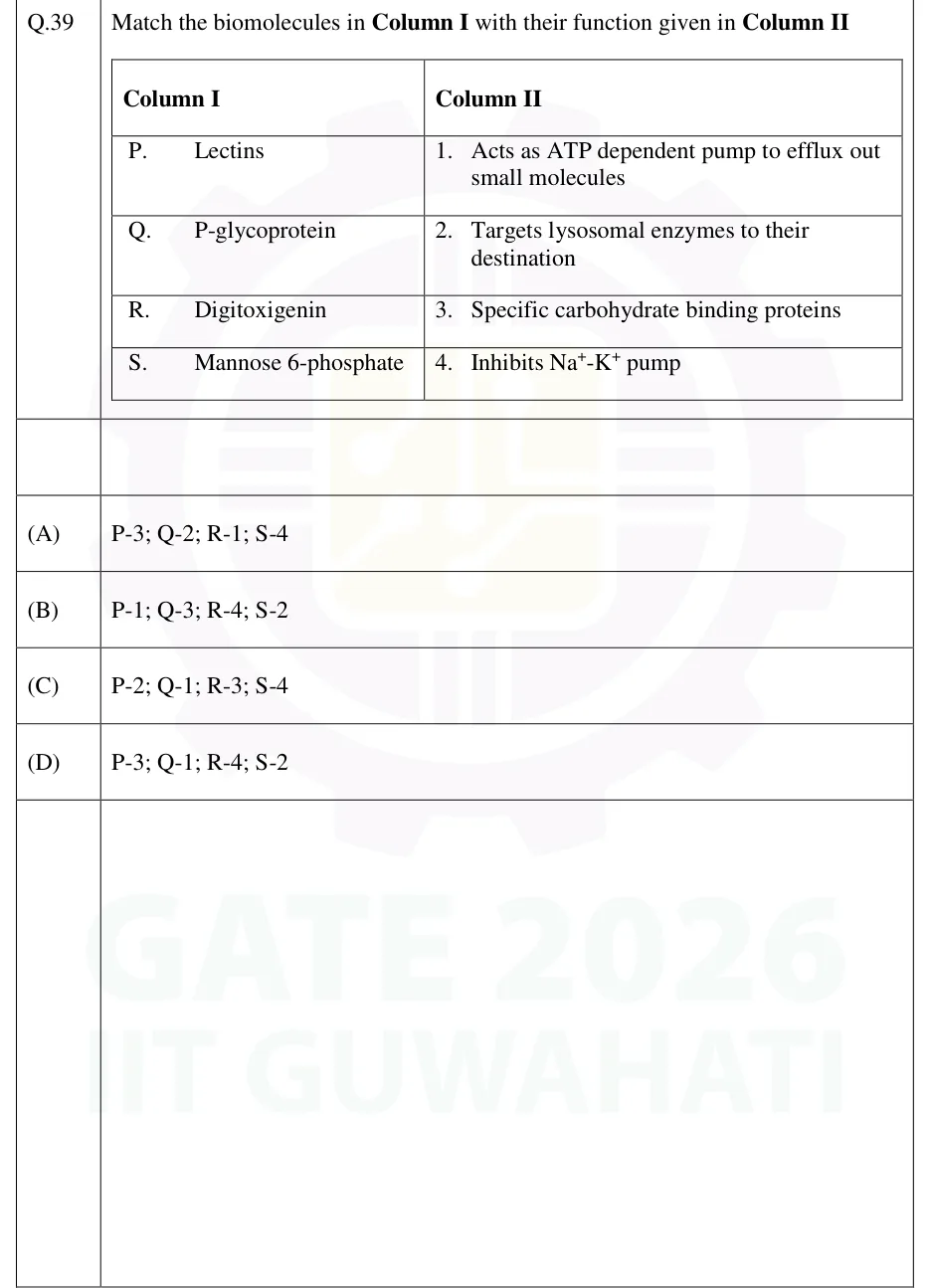
Match the biomolecules in Column I with their function given in Column II.

GATE 2026 BT question 39: A two-column table.Tap to open full size
Figure from the GATE 2026 BT paper. It is part of the question.
Figure description

A two-column table. Column I: P. Lectins; Q. P-glycoprotein; R. Digitoxigenin; S. Mannose 6-phosphate. Column II: 1. Acts as ATP dependent pump to efflux out small molecules; 2. Targets lysosomal enzymes to their destination; 3. Specific carbohydrate binding proteins; 4. Inhibits Na+-K+ pump.

  1. (A)

    P-3; Q-2; R-1; S-4

  2. (B)

    P-1; Q-3; R-4; S-2

  3. (C)

    P-2; Q-1; R-3; S-4

  4. (D)

    P-3; Q-1; R-4; S-2

Answer and solution
GATE 2026 BT Q.39: answer and solution

GATE 2025 BT Q.18

GATE 2025 · BTQ.18 · 1 mark · MCQ

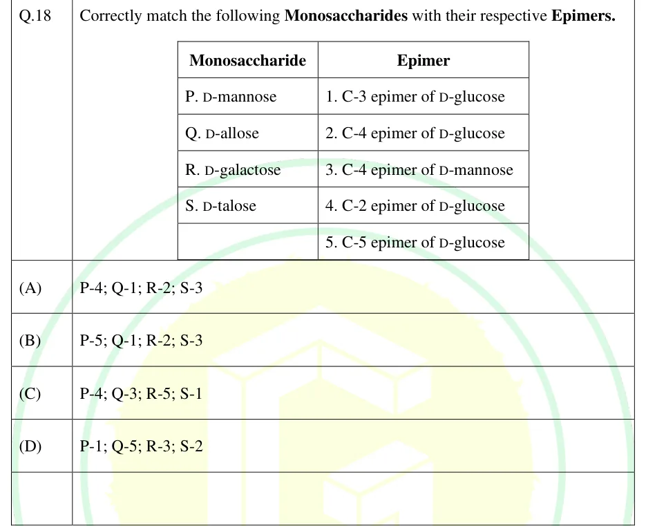
Correctly match the following Monosaccharides with their respective Epimers. Monosaccharide: P. D-mannose; Q. D-allose; R. D-galactose; S. D-talose. Epimer: 1. C-3 epimer of D-glucose; 2. C-4 epimer of D-glucose; 3. C-4 epimer of D-mannose; 4. C-2 epimer of D-glucose; 5. C-5 epimer of D-glucose.

GATE 2025 BT question 18: A two-column table.Tap to open full size
Figure from the GATE 2025 BT paper. It is part of the question.
Figure description

A two-column table. Left column header 'Monosaccharide' with rows P. D-mannose, Q. D-allose, R. D-galactose, S. D-talose and a blank fifth row. Right column header 'Epimer' with rows 1. C-3 epimer of D-glucose, 2. C-4 epimer of D-glucose, 3. C-4 epimer of D-mannose, 4. C-2 epimer of D-glucose, 5. C-5 epimer of D-glucose.

  1. (A)

    P-4; Q-1; R-2; S-3

  2. (B)

    P-5; Q-1; R-2; S-3

  3. (C)

    P-4; Q-3; R-5; S-1

  4. (D)

    P-1; Q-5; R-3; S-2

Answer and solution
GATE 2025 BT Q.18: answer and solution

GATE 2025 BT Q.38

GATE 2025 · BTQ.38 · 2 marks · MCQ

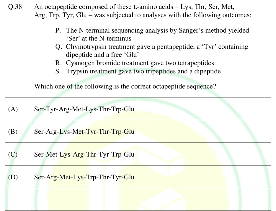
An octapeptide composed of these L-amino acids – Lys, Thr, Ser, Met, Arg, Trp, Tyr, Glu – was subjected to analyses with the following outcomes:
P. The N-terminal sequencing analysis by Sanger's method yielded 'Ser' at the N-terminus
Q. Chymotrypsin treatment gave a pentapeptide, a 'Tyr' containing dipeptide and a free 'Glu'
R. Cyanogen bromide treatment gave two tetrapeptides
S. Trypsin treatment gave two tripeptides and a dipeptide
Which one of the following is the correct octapeptide sequence?

  1. (A)

    Ser-Tyr-Arg-Met-Lys-Thr-Trp-Glu

  2. (B)

    Ser-Arg-Lys-Met-Tyr-Thr-Trp-Glu

  3. (C)

    Ser-Met-Lys-Arg-Thr-Tyr-Trp-Glu

  4. (D)

    Ser-Arg-Met-Lys-Trp-Thr-Tyr-Glu

Answer and solution
See the question in the paper
GATE 2025 BT Q.38: answer and solution

GATE 2023 BT Q.50

GATE 2023 · BTQ.50 · 2 marks · NAT

E. coli cultivated at 298 K uptakes an uncharged compound (A) by passive diffusion. The intracellular and extracellular concentrations of A are 0.001 M and 0.1 M, respectively. If the value of the ideal gas constant R is 1.9872 cal.mol^-1.K^-1, the free-energy change (in kcal.mol^-1) for this passive diffusion of A (rounded off to two decimal places) is ______.

Answer and solution
See the question in the paper
GATE 2023 BT Q.50: answer and solution

GATE 2022 BT Q.42

GATE 2022 · BTQ.42 · 2 marks · MCQ

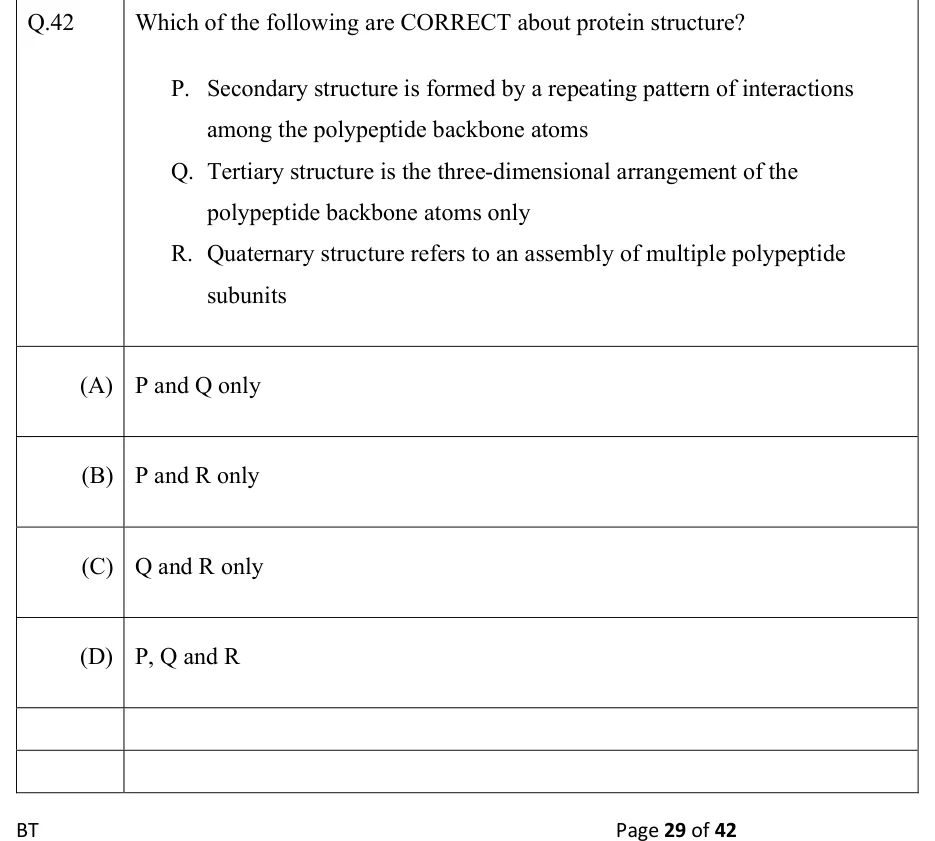
Which of the following are CORRECT about protein structure?
P. Secondary structure is formed by a repeating pattern of interactions among the polypeptide backbone atoms
Q. Tertiary structure is the three-dimensional arrangement of the polypeptide backbone atoms only
R. Quaternary structure refers to an assembly of multiple polypeptide subunits

  1. (A)

    P and Q only

  2. (B)

    P and R only

  3. (C)

    Q and R only

  4. (D)

    P, Q and R

Answer and solution
See the question in the paper
GATE 2022 BT Q.42: answer and solution

GATE 2019 BT Q.46

GATE 2019 · BTQ.46 · 2 marks · NAT

The difference in concentrations of an uncharged solute between two compartments is 1.6-fold. The energy required for active transport of the solute across the membrane separating the two compartments is ____ cal mol^-1 (rounded off to the nearest integer). (R = 1.987 cal mol^-1 K^-1, T = 37 °C)

Answer and solution
See the question in the paper
GATE 2019 BT Q.46: answer and solution

PRACTICE

6 questions to test yourself

PRACTICE 12 marks · NAT

Aspartic acid has pKa values of 1.99 (alpha-COOH), 3.90 (side-chain COOH) and 9.90 (alpha-NH3+). Calculate its isoelectric point (pI). Give the answer to two decimal places.

Answer and solution

Answer: (accepted range )

  1. At very low pH the net charge is +1 (both carboxyls neutral, amino +1).

  2. After pKa 1.99: net 0 (alpha-carboxylate -1, amino +1, side-chain carboxyl still neutral).

  3. After pKa 3.90: net -1.

  4. The zero-charge species lies between pKa 1.99 and 3.90.

  5. pI = (1.99 + 3.90)/2 = 2.945, about 2.95.

PRACTICE 22 marks · NAT

For a cell at 37 degrees C, the K+ concentration is 4 mM outside and 120 mM inside. Using the Nernst equation in the form E = (61.5 mV / z) log10([out]/[in]), calculate the equilibrium potential of K+ in mV. Give the answer to the nearest 0.1 mV (the answer is negative).

Answer and solution

Answer: (accepted range )

  1. z = +1 for K+.

  2. [out]/[in] = 4/120 = 1/30.

  3. log10(1/30) = -1.4771.

  4. E_K = 61.5 x (-1.4771) = -90.84 mV, about -90.8 mV.

PRACTICE 32 marks · MSQ

Which of the following statements about the Na+/K+-ATPase of animal cells are correct?

  1. (A)

    It moves 3 Na+ out of the cell and 2 K+ into the cell for each ATP hydrolysed.

  2. (B)

    It is a secondary active transporter driven by the Na+ gradient.

  3. (C)

    Its catalytic cycle includes a phosphorylated aspartate intermediate.

  4. (D)

    It is inhibited by ouabain.

Answer and solution

Answer: (A), (C), (D)

  1. A is correct: 3 Na+ out and 2 K+ in per ATP.

  2. B is wrong: it is a primary active transporter that uses ATP directly; it creates the Na+ gradient that secondary transporters use.

  3. C is correct: it is a P-type ATPase, phosphorylated on an aspartate residue.

  4. D is correct: ouabain is a classic inhibitor.

PRACTICE 41 mark · MCQ

In an axon, which motor protein moves cargo along microtubules toward the minus end, bringing material from the axon terminal back toward the cell body?

  1. (A)

    Conventional kinesin

  2. (B)

    Cytoplasmic dynein

  3. (C)

    Myosin II

  4. (D)

    Myosin V

Answer and solution

Answer: (B)

  1. In an axon, microtubule minus ends point towards the cell body and plus ends towards the terminal.

  2. Conventional kinesin moves to the plus end (towards the terminal): anterograde.

  3. Cytoplasmic dynein moves to the minus end: retrograde transport.

  4. Myosins move on actin, not microtubules.

PRACTICE 52 marks · MSQ

Which of the following statements about storage polysaccharides are correct?

  1. (A)

    A glycogen molecule has many non-reducing ends but only one reducing end.

  2. (B)

    Amylopectin contains alpha(1-6) branch points.

  3. (C)

    Glycogen is less highly branched than amylopectin.

  4. (D)

    Amylose chains are joined by beta(1-4) glycosidic bonds.

Answer and solution

Answer: (A), (B)

  1. A is correct: every branch ends in a non-reducing end (free C4-OH); only the one glucose whose anomeric carbon is not in a bond is a reducing end.

  2. B is correct: amylopectin has alpha(1-4) chains with alpha(1-6) branch points.

  3. C is wrong: glycogen branches about every 8 to 12 residues, more often than amylopectin (about every 24 to 30).

  4. D is wrong: amylose is alpha(1-4); beta(1-4) glucose links are found in cellulose.

PRACTICE 61 mark · MCQ

Which of the following disaccharides is NOT a reducing sugar?

  1. (A)

    Maltose

  2. (B)

    Lactose

  3. (C)

    Sucrose

  4. (D)

    Cellobiose

Answer and solution

Answer: (C)

  1. A reducing sugar needs a free anomeric carbon that can open to the carbonyl form.

  2. Maltose, lactose and cellobiose each have one glucose whose anomeric carbon is not in the linkage, so they are reducing.

  3. In sucrose the glycosidic bond joins C1 of glucose to C2 of fructose, and both are anomeric carbons.

  4. So sucrose is non-reducing.

OFFICIAL GATE 2027 SYLLABUS WORDING

Copied from the GATE 2027 syllabus; see all 2027 syllabi.

GATE BT

Biochemistry: Biomolecules - structure and function; Biological membranes - structure, membrane channels and pumps, molecular motors, action potential and transport processes

Preparing in Kolkata? IAE Academy's course for GATE BT.