IAE Academy

GATE BT

Pathogens & antibiotics

Operons, nitrogen fixation, pathogens, antibiotics and viruses

This topic covers how bacteria switch genes on and off (lac, trp and ara operons), how nitrogen is fixed, how microbes cause disease, how antibiotics work and resistance arises, how cells sense signals (two-component systems, quorum sensing), and how viruses are built and classified. The notes focus on reasoning through operon mutants and partial diploids, matching toxins, drugs and viruses to their targets, and placing viruses in Baltimore classes, so a mechanism-first understanding pays off.

11 previous-year questions · 11 solved · 6 practice questions

The lac operon

The lac operon of E. coli lets the cell use lactose only when it is present and glucose is scarce. It has two levels of control: a negative one (the repressor) and a positive one (CAP-cAMP). Convention used here: gene names are written plainly in lower case (lacZ, lacY, lacA, lacI) and protein names with a capital letter (LacI, TrpR, AraC).

Element

Product / role

lacI

Repressor protein LacI (tetramer); made constitutively from its own weak promoter

lacP

Promoter of the structural genes

lacO

Operator; the repressor binds here and blocks transcription by RNA polymerase

lacZ

β-galactosidase; splits lactose into glucose and galactose and also converts some lactose to allolactose

lacY

Lactose permease; membrane transporter that takes lactose in

lacA

Thiogalactoside transacetylase

CAP site

Binding site upstream of the promoter for the CAP-cAMP complex (positive control)

Negative control. With no lactose, the repressor sits on the operator and the operon is off (a very low basal level remains). When lactose enters, the few β-galactosidase molecules made at the basal level convert some of it to allolactose, the natural inducer, which binds the repressor, changes its shape and releases it from the operator. IPTG is a gratuitous inducer: it induces but is not metabolised.

Positive control and catabolite repression. Low glucose raises cAMP. The CAP (also called CRP) protein binds cAMP, and the CAP-cAMP complex binds near the promoter and helps RNA polymerase start. High glucose lowers cAMP, so CAP is inactive and the operon stays at a low level even if lactose is present. Glucose also acts by inducer exclusion: while glucose is taken up by the phosphotransferase system, the unphosphorylated EIIA(Glc) protein binds LacY and blocks lactose entry. Maximal expression therefore needs both lactose present (repressor off) and glucose absent (CAP-cAMP on).

Glucose

Lactose

Repressor

CAP-cAMP

Expression

+

-

On operator

Inactive

Off (basal level only)

+

+

Off operator

Inactive

Low

-

-

On operator

Active

Off (basal level only)

-

+

Off operator

Active

High

Mutation

Phenotype

Cis or trans?

lacI⁻ (no functional repressor)

Constitutive expression; recessive to lacI⁺

Acts in trans (diffusible protein)

lacIˢ (super-repressor; cannot bind inducer)

Never induced; dominant over lacI⁺

Acts in trans

lacI⁻ᵈ (dominant-negative repressor)

Mutant subunits form mixed tetramers with wild-type subunits that cannot bind the operator; constitutive even with lacI⁺ present

Acts in trans

lacOᶜ (operator cannot bind repressor)

Constitutive expression of the genes on the same DNA only; dominant in cis

Cis-acting only

lacP⁻ (promoter defect)

No transcription of the genes on the same DNA

Cis-acting only

lacZ⁻ or lacY⁻

Loss of that enzyme; recessive, complemented by the wild-type allele on the other copy

Trans (protein products)

Procedure for partial-diploid (merodiploid) questions

  1. Write each copy of the DNA separately: its lacI, lacP, lacO and structural genes.

  2. Decide which repressor is present in the cell. A repressor made by any lacI⁺ copy acts on every lacO⁺ operator, on either copy. lacIˢ is dominant and cannot be released by inducer; lacI⁻ᵈ poisons wild-type subunits; lacI⁻ makes nothing.

  3. A copy is transcribed only if its own promoter works and its own operator is free: either the operator cannot bind repressor (lacOᶜ), or no functional repressor is present, or inducer has released the repressor (not possible with lacIˢ).

  4. Then check whether that same copy carries a functional lacZ or lacY; operator and promoter defects act only on their own copy.

  5. Repeat for each condition asked. A high level also needs CAP-cAMP, that is, glucose absent.

EXAM TIP

Trans-acting: the repressor (lacI) and the enzymes. Cis-acting: the operator and promoter. lacOᶜ makes only the genes on its own DNA constitutive, so in lacI⁺ lacOᶜ lacZ⁻ / lacI⁺ lacO⁺ lacZ⁺ the constitutive copy makes a defective enzyme and the normal copy stays inducible.

WORKED EXAMPLE

A lac merodiploid is F' lacI⁺ lacOᶜ lacZ⁻ / chromosome lacI⁺ lacO⁺ lacZ⁺. Is functional β-galactosidase made without lactose?

Solution
  1. The chromosome copy has a normal operator, bound by repressor from lacI⁺, so it is off when there is no inducer.

  2. The F' copy has lacOᶜ, so its operator cannot bind repressor and it is transcribed, but its lacZ is defective.

  3. The only functional lacZ is on the chromosome, and it is repressed.

Answer: No functional β-galactosidase is made without lactose (beyond the basal level). With lactose, the chromosomal copy is induced and functional enzyme appears.

The trp and ara operons

The trp operon encodes five polypeptides (trpE, trpD, trpC, trpB, trpA) that form the enzymes building tryptophan from chorismate, among them anthranilate synthase (TrpE with TrpD) and tryptophan synthase (TrpA with TrpB). It is a repressible operon: the end product switches it off. Two layers of control act: repression and attenuation.

Repression. TrpR is made as an inactive aporepressor. When tryptophan is abundant it acts as a corepressor: it binds TrpR, which then binds the operator and blocks transcription. When tryptophan is scarce, it dissociates from TrpR, the repressor leaves the operator and transcription starts.

Attenuation. A leader region (trpL) lies between the operator and trpE. It has four stretches (1, 2, 3, 4), and region 1 encodes a short leader peptide with two adjacent tryptophan codons. The RNA can pair 1-2, 2-3 or 3-4. The 3-4 hairpin followed by a run of U's is a rho-independent terminator; the 2-3 hairpin is an anti-terminator, and it prevents 3-4 from forming. Because transcription and translation are coupled in bacteria, the ribosome's position on the leader decides which hairpin forms. The ribosome stalls at the Trp codons when charged tRNA(Trp) is scarce. If no ribosome translates the leader at all, 1-2 and 3-4 form and transcription terminates.

Tryptophan level

Ribosome behaviour on leader

Hairpin formed

Result

High

Translates region 1 quickly and covers region 2

3-4 terminator

Transcription stops before trpE (attenuated)

Low

Stalls at the Trp codons in region 1

2-3 anti-terminator

Transcription continues into trpE

EXAM TIP

Repression and attenuation act independently. A repressor-defective (trpR⁻) strain is still regulated by tryptophan through attenuation, and a strain whose leader cannot attenuate is still regulated by repression.

The ara operon controls arabinose use. araBAD encodes ribulokinase (araB), L-arabinose isomerase (araA) and L-ribulose-5-phosphate 4-epimerase (araD). The regulator AraC is both a repressor and an activator. Without arabinose, an AraC dimer binds the araO2 and araI1 sites, which are far apart, and loops the DNA, which represses. With arabinose, AraC binds arabinose, changes conformation, releases araO2 and binds the adjacent sites araI1 and araI2, which activates transcription from the araBAD promoter. Activation also needs CAP-cAMP, so the ara operon, like lac, is subject to catabolite repression. AraC also represses its own gene, araC, by binding araO1.

Feature

lac

trp

ara

Type

Inducible

Repressible

Inducible

Small molecule

Allolactose (inducer)

Tryptophan (corepressor); attenuation senses the level of charged tRNA(Trp)

Arabinose (inducer; changes AraC)

Regulator

LacI repressor (negative)

TrpR repressor (negative) plus attenuation

AraC (positive and negative)

CAP-cAMP

Required for activation

Not involved

Required for activation

Nitrogen fixation

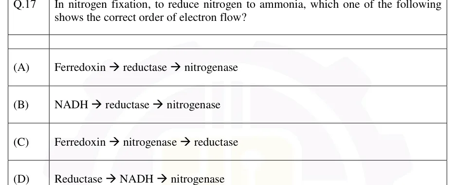
Nitrogen fixation is the reduction of atmospheric N₂ to ammonia. Only certain prokaryotes (bacteria and archaea) carry it out. The enzyme is nitrogenase, which has two components: the Fe protein (dinitrogenase reductase, encoded by nifH) and the MoFe protein (dinitrogenase, encoded by nifD and nifK). The Fe protein, driven by ATP hydrolysis, passes electrons one at a time to the MoFe protein, whose iron-molybdenum cofactor (FeMo-co) binds and reduces N₂. Both components are irreversibly damaged by oxygen.

NITROGENASE REACTION (CONVENTIONAL STOICHIOMETRY)

Convention: 2 ATP hydrolysed per electron transferred; 8 electrons per N₂ (6 to make 2 NH₃ and 2 for the obligatory H₂), so 16 ATP per N₂. In cells the cost is often higher because extra H₂ is made.

Group

Examples

Notes

Symbiotic, legume nodules

Rhizobium, Bradyrhizobium, Sinorhizobium

Nodules with leghemoglobin; nod genes for nodulation; host supplies carbon

Symbiotic, non-legume

Frankia (actinorhizal plants such as Alnus)

Filamentous actinobacterium

Symbiotic, cyanobacterial

Anabaena with Azolla

Fixation in heterocysts

Free-living aerobes

Azotobacter

Protects nitrogenase by a very high respiration rate and slime

Free-living anaerobes

Clostridium; Klebsiella (facultative, fixes only without O₂)

Fixation in the absence of O₂

Free-living phototrophs

Cyanobacteria such as Nostoc, Anabaena

Heterocysts separate fixation from oxygenic photosynthesis

Associative

Azospirillum

Lives around the roots of grasses

Protecting nitrogenase from oxygen. Leghemoglobin in the nodule (the globin is made by the plant) binds O₂ tightly, keeping free O₂ low while still supplying enough for bacteroid respiration. Heterocysts have thick walls and lack the oxygen-evolving photosystem II. Azotobacter respires rapidly. The nif genes (nifH, nifD, nifK, and regulators such as nifA) are switched off by high ammonia and by oxygen.

EXAM TIP

The ammonia produced is assimilated into glutamine by glutamine synthetase, and glutamate is made by GOGAT (glutamine + 2-oxoglutarate → 2 glutamate). In nodulation, flavonoids from the plant root activate the bacterial NodD protein, which switches on the nod genes; these make Nod factors (lipochitooligosaccharides) that trigger nodule formation. Some bacteria, such as Azotobacter, also have alternative nitrogenases with vanadium or only iron in place of molybdenum. Nitrogenase also reduces acetylene to ethylene, which is the basis of the acetylene reduction assay for nitrogenase activity.

Microbial disease and host-pathogen interactions

A pathogen causes disease through steps: adherence and colonisation, invasion, evasion of host defences, damage to tissue (often by toxins), and spread. Virulence factors are the molecules that make these steps possible: adhesins and pili, capsules (resist phagocytosis), secretion systems, iron-scavenging siderophores, enzymes (hyaluronidase, coagulase) and toxins. Many protein exotoxins are A-B toxins: the B part binds a receptor on the host cell and the A part is the active enzyme that enters the cell.

Two standard ideas

  1. Koch's postulates: the organism is found in every case of the disease; it is isolated and grown in pure culture; the pure culture causes the disease in a susceptible host; and the same organism is re-isolated from that host.

  2. Type III secretion system: a needle-like apparatus that injects bacterial effector proteins directly into host cells, used by gram-negative pathogens such as Salmonella, Shigella and Yersinia.

Property

Exotoxin

Endotoxin

Chemical nature

Protein

Lipopolysaccharide (the lipid A part is toxic)

Source

Made by both gram-positive and gram-negative bacteria; usually secreted by living cells

Part of the gram-negative outer membrane; released mainly on cell lysis

Potency / specificity

Very potent; specific effect on a host target

Less potent; general effects (fever, shock)

Heat stability

Mostly heat-labile

Heat-stable

Immunogenicity

Strongly antigenic; induces neutralising antitoxin

Weakly immunogenic

Toxoid formation

Can be converted into toxoids (vaccines)

Not converted to toxoids

Genes

Often on plasmids or phages

Chromosomal

Toxin or organism

Mechanism or disease

Cholera toxin (Vibrio cholerae)

ADP-ribosylates the α subunit of Gs, which then cannot hydrolyse GTP; adenylate cyclase stays active; high cAMP; watery diarrhoea

Pertussis toxin (Bordetella pertussis)

ADP-ribosylates the α subunit of Gi, which can no longer inhibit adenylate cyclase; cAMP rises

Diphtheria toxin (Corynebacterium diphtheriae)

ADP-ribosylates elongation factor 2 (EF-2); blocks host protein synthesis

Shiga toxin

Removes an adenine from 28S rRNA of the large ribosomal subunit; blocks protein synthesis

Tetanus toxin (Clostridium tetani)

Cleaves a SNARE protein in inhibitory neurons, blocking release of the inhibitory transmitters glycine and GABA; spastic paralysis

Botulinum toxin (Clostridium botulinum)

Cleaves SNARE proteins at the neuromuscular junction, blocking acetylcholine release; flaccid paralysis

Superantigens (staphylococcal enterotoxins, toxic shock syndrome toxin-1)

Bind MHC class II and the T-cell receptor outside the peptide groove, activating many T cells at once; massive cytokine release

Anthrax (Bacillus anthracis)

Toxin of three proteins: protective antigen (the binding part), lethal factor (a protease) and edema factor (an adenylate cyclase); capsule of poly-D-glutamate

Tuberculosis (Mycobacterium tuberculosis)

Survives inside macrophages by blocking phagosome-lysosome fusion; granulomas; acid-fast cell wall rich in mycolic acids

Typhoid (Salmonella Typhi), plague (Yersinia pestis), leprosy (Mycobacterium leprae), syphilis (Treponema pallidum)

Classical bacterial diseases

Viral diseases

Polio (poliovirus), influenza (influenza virus), AIDS (HIV), hepatitis B (hepatitis B virus), rabies, measles, dengue, COVID-19 (SARS-CoV-2)

EXAM TIP

Several toxin genes are carried by phages: cholera toxin (phage CTXφ), diphtheria toxin (phage β) and the Shiga toxins of enterohaemorrhagic E. coli. Lysogenic conversion can therefore turn a harmless strain into a toxin-producing one.

Antibiotics and antimicrobial resistance

Antibiotics are selectively toxic: they hit targets that bacteria have and human cells lack or have in a different form (for example 70S versus 80S ribosomes). Bactericidal drugs kill bacteria; bacteriostatic drugs stop growth and rely on the host immune system to clear the infection. The minimum inhibitory concentration (MIC) is the lowest drug concentration that prevents visible growth in a defined culture after overnight incubation; the minimum bactericidal concentration (MBC) is the lowest that kills at least 99.9% of the cells.

Target

Antibiotic

Action

Usual effect

Cell wall

Penicillins, cephalosporins (β-lactams)

Inhibit the transpeptidases (penicillin-binding proteins) that cross-link peptidoglycan

Bactericidal

Cell wall

Vancomycin

Binds the D-Ala-D-Ala terminus of the peptidoglycan precursor

Bactericidal

Cell wall (mycobacteria)

Isoniazid

Prodrug activated by KatG; inhibits mycolic acid synthesis (InhA)

Bactericidal against dividing mycobacteria

Protein synthesis, 30S

Streptomycin, gentamicin (aminoglycosides)

Cause misreading of mRNA

Bactericidal

Protein synthesis, 30S

Tetracycline

Blocks aminoacyl-tRNA binding to the A site

Bacteriostatic

Protein synthesis, 50S

Chloramphenicol

Inhibits peptidyl transferase

Bacteriostatic

Protein synthesis, 50S

Macrolides (erythromycin), clindamycin

Bind near the peptidyl transferase centre and the exit tunnel; block chain elongation

Bacteriostatic

Protein synthesis (research use)

Puromycin

Aminoacyl-tRNA analogue; accepted at the A site and causes premature chain release; acts on both bacterial and eukaryotic ribosomes

Not used clinically

Nucleic acids

Rifampicin

Inhibits bacterial RNA polymerase (β subunit)

Bactericidal

Nucleic acids

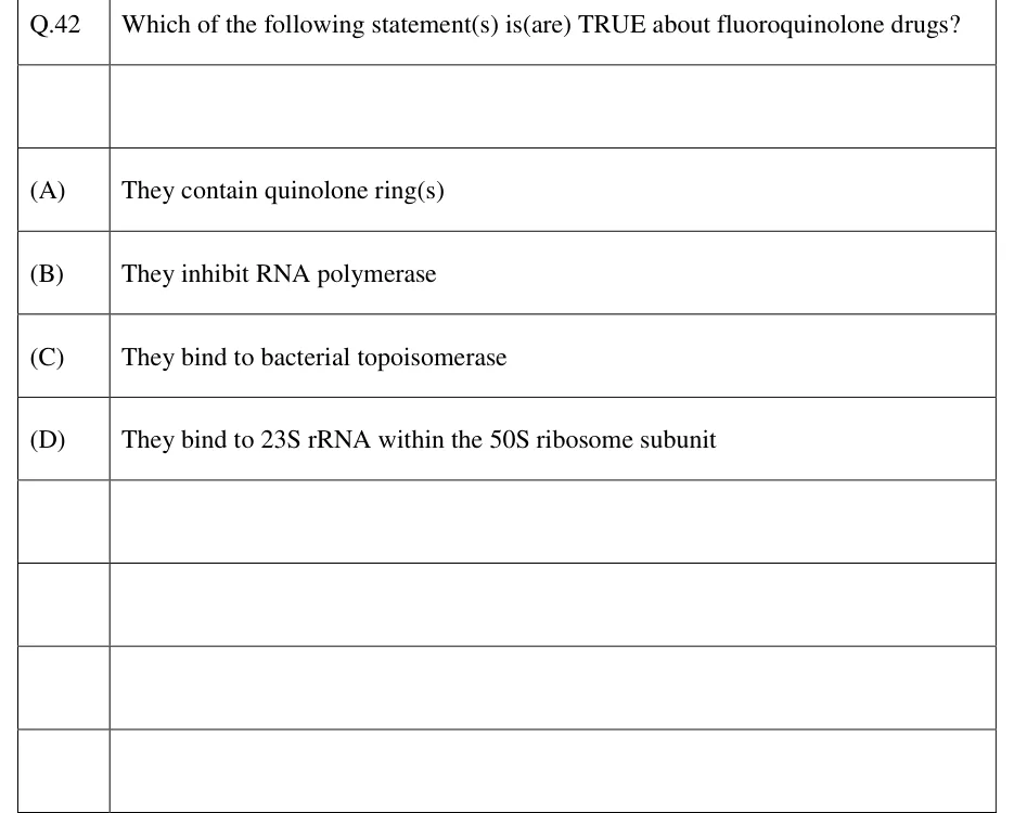
Fluoroquinolones (ciprofloxacin)

Inhibit DNA gyrase and topoisomerase IV

Bactericidal

Folate synthesis

Sulfonamides; trimethoprim

Sulfonamides inhibit dihydropteroate synthase; trimethoprim inhibits dihydrofolate reductase

Each bacteriostatic alone; the combination is usually bactericidal

Membrane

Polymyxin (gram-negative, binds LPS); daptomycin (gram-positive)

Disrupt membrane integrity

Bactericidal

Resistance mechanism

Example

Enzymatic inactivation of the drug

β-lactamases cleave penicillins; aminoglycoside-modifying enzymes; chloramphenicol acetyltransferase

Alteration of the target

mecA gives PBP2a in MRSA; D-Ala-D-Lac precursors in vancomycin resistance; methylation of 23S rRNA (erm) against macrolides; gyrase mutations against fluoroquinolones

Efflux pumps

Tetracycline efflux (Tet pumps); multidrug pumps

Reduced permeability

Loss or change of porins in the gram-negative outer membrane

Bypass pathway

Alternative enzyme not inhibited by the drug (for example trimethoprim-resistant dihydrofolate reductase)

Resistance spreads vertically by mutation and inheritance, and horizontally by conjugation (R plasmids), transformation and transduction, with transposons and integrons moving resistance genes and cassettes between replicons. Antibiotic use selects for resistant cells. Clavulanic acid, a β-lactamase inhibitor, is combined with amoxicillin to protect it.

EXAM TIP

Gram-negative bacteria are intrinsically less permeable to many drugs because of their outer membrane. Vancomycin, a large molecule, does not cross it and so works against gram-positive bacteria. Cycloheximide inhibits translation on eukaryotic 80S ribosomes but not on bacterial 70S ribosomes, whereas puromycin acts on both.

Two-component systems and quorum sensing

A two-component system (TCS) is the usual way bacteria sense the environment. A sensor histidine kinase, usually a membrane protein, detects a signal and autophosphorylates a conserved histidine using ATP. The phosphoryl group is then transferred to a conserved aspartate on a cytoplasmic response regulator. Phosphorylation activates the regulator, which is often a transcription factor. The system resets when the phosphate is removed, either by the regulator's own slow dephosphorylation or by a phosphatase activity, often of the sensor itself.

System

Signal / response

EnvZ-OmpR

Osmolarity; changes porin (ompF, ompC) expression

NtrB-NtrC

Nitrogen limitation; activates nitrogen assimilation genes

PhoQ-PhoP

Low Mg²⁺; virulence control in Salmonella

CheA-CheY

Chemotaxis; CheA is a cytoplasmic kinase coupled to the chemoreceptors; phosphorylated CheY binds the flagellar motor and causes tumbling

VanS-VanR

Vancomycin; induces resistance genes

Quorum sensing lets cells coordinate behaviour by measuring cell density through secreted signals called autoinducers. When the signal concentration crosses a threshold, a group of genes is switched on together. Targets include bioluminescence, virulence factors and biofilm formation.

Feature

Gram-negative (Vibrio fischeri model)

Gram-positive (for example Staphylococcus aureus agr)

Signal

Acyl-homoserine lactone (AHL), small and diffusible

Short processed oligopeptide (autoinducing peptide)

Synthase

LuxI

Peptide precursor processed and exported

Detector

LuxR, an intracellular receptor and transcription factor

Membrane sensor kinase of a two-component system

Output

AHL-LuxR complex activates the luciferase (lux) genes

Phosphorylated response regulator switches on target genes

EXAM TIP

Autoinducer-2 is a signal used between different species. It is derived from DPD, the product of the LuxS enzyme; in Vibrio harveyi it is a furanosyl borate diester. Quorum sensing often includes positive feedback (in V. fischeri the AHL-LuxR complex also induces luxI), which gives an abrupt switch. His-Asp phosphorelay systems are found in bacteria, archaea, fungi and plants (for example the ethylene and cytokinin receptors), but not in animals.

Viruses: structure, classification and replication

A virus is an obligate intracellular particle whose genome is either DNA or RNA, not both, inside a protein shell called the capsid, built from capsomers. The capsid plus genome is the nucleocapsid. Some viruses also have a lipid envelope taken from a host membrane, carrying viral glycoproteins. Capsid symmetry is helical (tobacco mosaic virus), icosahedral (adenovirus, poliovirus) or complex (T4 phage, poxviruses). Viruses lack their own ribosomes and metabolism. Taxonomic classification uses the nature of the genome, capsid symmetry, presence of an envelope and size; the Baltimore scheme groups viruses by how they make mRNA.

Baltimore class

Genome

Examples

Key feature

I

dsDNA

Herpesviruses, adenovirus, T4 phage, poxvirus

mRNA made from the DNA template

II

ssDNA

Parvovirus, M13 and ϕX174 phages

Converted to dsDNA before transcription

III

dsRNA

Rotavirus, reovirus

Virus carries its own RNA-dependent RNA polymerase

IV

ssRNA(+)

Poliovirus, hepatitis C virus, SARS-CoV-2, TMV

Genome acts directly as mRNA

V

ssRNA(-)

Influenza virus, measles virus, rabies virus

Needs a virion RNA-dependent RNA polymerase to make mRNA

VI

ssRNA(+) with reverse transcriptase

HIV (retroviruses)

Diploid positive-sense RNA genome; reverse transcriptase makes DNA, which integrates into the host genome

VII

dsDNA (partly double-stranded) with reverse transcription

Hepatitis B virus

Replicates through an RNA intermediate (pregenomic RNA) copied by reverse transcriptase

Subviral agents. Viroids are small, circular, single-stranded RNA molecules with no protein coat and no protein-coding genes that infect plants (for example potato spindle tuber viroid). Prions are infectious proteins with no nucleic acid: a misfolded form of the prion protein (PrP) converts the normal form to the misfolded form, causing spongiform encephalopathies.

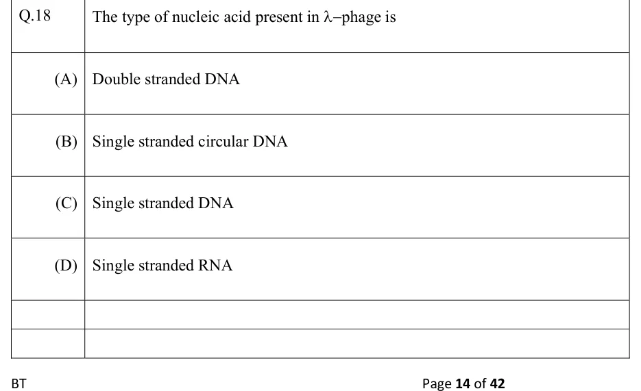
Bacteriophage cycles. In the lytic cycle a virulent phage adsorbs, injects its DNA, directs the synthesis of new phage, assembles and lyses the cell. In the lysogenic cycle a temperate phage such as lambda integrates as a prophage and replicates with the host. In lambda, the cI repressor maintains lysogeny. DNA damage triggers the SOS response: activated RecA (RecA*) stimulates self-cleavage of cI, and the phage enters the lytic cycle (induction).

One-step growth curve. After a synchronous infection, the eclipse period is the time during which no infectious phage can be recovered even from broken cells; the latent period lasts until the cells lyse and release phage. The burst size is the average number of progeny phage released per infected cell.

Transduction is gene transfer by phage. In generalised transduction (for example phage P1), random host DNA is packaged by mistake, so any gene can be transferred. In specialised transduction, a prophage that excises imprecisely carries only the host genes next to its integration site, as with lambda carrying gal or bio.

PLAQUE ASSAY TITER

Use a plate with a countable number of plaques (conventionally 30 to 300). The dilution is the fraction of the original stock in the plated sample (such as ).

MULTIPLICITY OF INFECTION (POISSON DISTRIBUTION)

m is the multiplicity of infection (phage added per bacterium), assuming random adsorption. P(k) is the fraction of cells infected by exactly k phages.

WORKED EXAMPLE

A phage stock is diluted and 0.2 mL is plated. It gives 60 plaques. Find the titer of the original stock.

Solution
  1. Use titer = plaques / (dilution × volume).

  2. Titer .

  3. Titer PFU/mL.

Answer: PFU/mL

PREVIOUS-YEAR QUESTIONS

11 questions from the BT papers

Try each question first, then open the answer. We placed each question under a topic ourselves; the exam paper does not label topics.

See these papers in full: GATE BT 2026 · GATE BT 2024 · GATE BT 2023 · GATE BT 2022 · GATE BT 2020 · GATE BT 2019

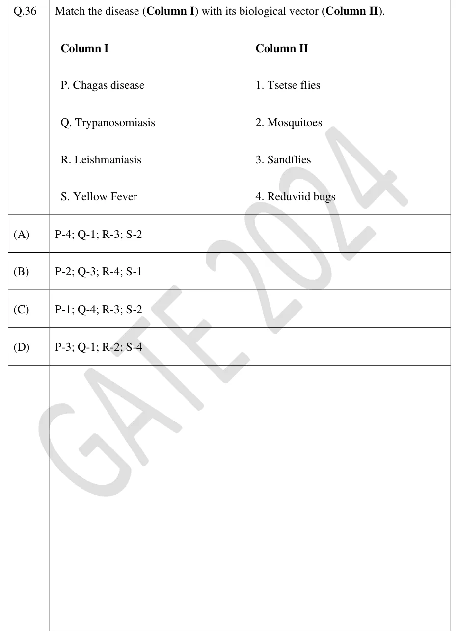
GATE 2024 BT Q.36

GATE 2024 · BTQ.36 · 2 marks · MCQ

Match the disease (Column I) with its biological vector (Column II).

Column I: P. Chagas disease; Q. Trypanosomiasis; R. Leishmaniasis; S. Yellow Fever
Column II: 1. Tsetse flies; 2. Mosquitoes; 3. Sandflies; 4. Reduviid bugs

  1. (A)

    P-4; Q-1; R-3; S-2

  2. (B)

    P-2; Q-3; R-4; S-1

  3. (C)

    P-1; Q-4; R-3; S-2

  4. (D)

    P-3; Q-1; R-2; S-4

Answer and solution
See the question in the paper
GATE 2024 BT Q.36: answer and solution

GATE 2023 BT Q.20

GATE 2023 · BTQ.20 · 1 mark · MCQ

The correct sequence of events during bacteriophage infection of a bacterial cell is

  1. (A)

    landing → attachment → tail contraction → penetration and unplugging → DNA ejection

  2. (B)

    attachment → landing → penetration and unplugging → tail contraction → DNA ejection

  3. (C)

    landing → tail contraction → attachment → DNA ejection → penetration and unplugging

  4. (D)

    attachment → tail contraction → landing → penetration and unplugging → DNA ejection

Answer and solution
See the question in the paper
GATE 2023 BT Q.20: answer and solution

GATE 2023 BT Q.38

GATE 2023 · BTQ.38 · 2 marks · MCQ

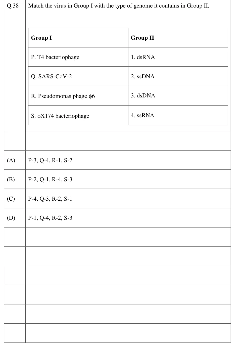
Match the virus in Group I with the type of genome it contains in Group II.

Group I: P. T4 bacteriophage; Q. SARS-CoV-2; R. Pseudomonas phage φ6; S. φX174 bacteriophage.
Group II: 1. dsRNA; 2. ssDNA; 3. dsDNA; 4. ssRNA.

  1. (A)

    P-3, Q-4, R-1, S-2

  2. (B)

    P-2, Q-1, R-4, S-3

  3. (C)

    P-4, Q-3, R-2, S-1

  4. (D)

    P-1, Q-4, R-2, S-3

Answer and solution
See the question in the paper
GATE 2023 BT Q.38: answer and solution

GATE 2020 BT Q.34

GATE 2020 · BTQ.34 · 2 marks · MCQ

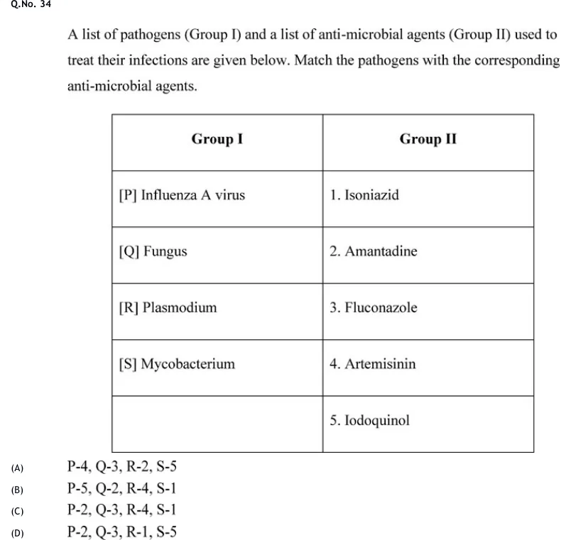
A list of pathogens (Group I) and a list of anti-microbial agents (Group II) used to treat their infections are given below. Match the pathogens with the corresponding anti-microbial agents.

GATE 2020 BT question 34: A two-column table.Tap to open full size
Figure from the GATE 2020 BT paper. It is part of the question.
Figure description

A two-column table. Group I: [P] Influenza A virus; [Q] Fungus; [R] Plasmodium; [S] Mycobacterium (a fifth Group I cell is blank). Group II: 1. Isoniazid; 2. Amantadine; 3. Fluconazole; 4. Artemisinin; 5. Iodoquinol.

  1. (A)

    P-4, Q-3, R-2, S-5

  2. (B)

    P-5, Q-2, R-4, S-1

  3. (C)

    P-2, Q-3, R-4, S-1

  4. (D)

    P-2, Q-3, R-1, S-5

Answer and solution
GATE 2020 BT Q.34: answer and solution

GATE 2019 BT Q.7

GATE 2019 · BTQ.7 · 1 mark · MCQ

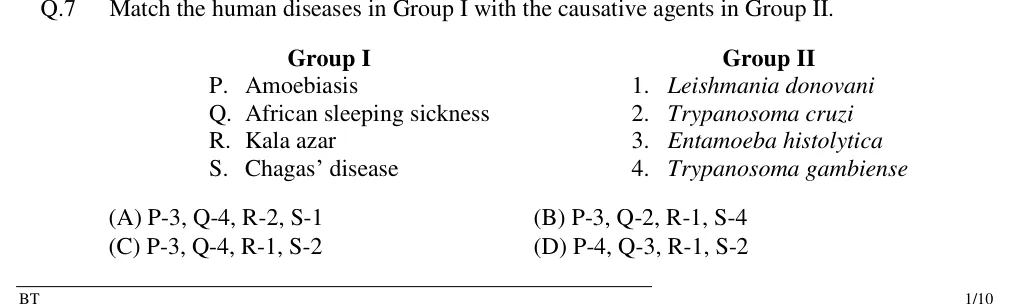
Match the human diseases in Group I with the causative agents in Group II.

Group I: P. Amoebiasis; Q. African sleeping sickness; R. Kala azar; S. Chagas' disease
Group II: 1. Leishmania donovani; 2. Trypanosoma cruzi; 3. Entamoeba histolytica; 4. Trypanosoma gambiense

  1. (A)

    P-3, Q-4, R-2, S-1

  2. (B)

    P-3, Q-2, R-1, S-4

  3. (C)

    P-3, Q-4, R-1, S-2

  4. (D)

    P-4, Q-3, R-1, S-2

Answer and solution
See the question in the paper
GATE 2019 BT Q.7: answer and solution

PRACTICE

6 questions to test yourself

PRACTICE 11 mark · MCQ

An E. coli merodiploid has the genotype F' lacI⁻ lacO⁺ lacZ⁺ / chromosome lacI⁺ lacO⁺ lacZ⁻. Cells are grown in the absence of glucose. Which statement is correct?

  1. (A)

    β-galactosidase is made with and without lactose

  2. (B)

    β-galactosidase is made only when lactose is present

  3. (C)

    β-galactosidase is never made

  4. (D)

    β-galactosidase is made only without lactose

Answer and solution

Answer: (B)

  1. lacI⁺ on the chromosome makes repressor, which acts in trans and can bind both operators (both are lacO⁺).

  2. Without lactose, both operators are bound, so neither copy is transcribed above the basal level.

  3. With lactose, allolactose releases the repressor, and the F' copy, which carries the functional lacZ, is expressed (CAP-cAMP is active because glucose is absent).

  4. So functional β-galactosidase appears only with lactose.

PRACTICE 21 mark · NAT

A phage lysate is diluted and 0.1 mL of the dilution is plated, giving 84 plaques. What is the titer of the lysate in units of PFU/mL? (Give the numerical value.)

Answer and solution

Answer: (accepted range )

  1. Titer = plaques / (dilution × volume).

  2. Titer .

  3. Titer PFU/mL, so the value in units of is 8.4.

PRACTICE 32 marks · NAT

Use the conventional nitrogenase stoichiometry: 2 ATP are hydrolysed per electron transferred, and 8 electrons are needed per N₂ reduced (6 to make 2 NH₃ and 2 for the obligatory H₂). How many moles of ATP are consumed to produce 6 mol of NH₃?

Answer and solution

Answer: (accepted range )

  1. Each N₂ gives 2 NH₃, so 6 mol NH₃ needs 3 mol N₂.

  2. Electrons: 3 mol N₂ × 8 = 24 mol electrons.

  3. ATP: 24 mol electrons × 2 = 48 mol ATP (equivalently 16 ATP per N₂ × 3).

PRACTICE 41 mark · MCQ

A virus packages a partly double-stranded DNA genome in its virion but copies its genome through an RNA intermediate using reverse transcriptase. Which virus is it?

  1. (A)

    HIV

  2. (B)

    Hepatitis B virus

  3. (C)

    Influenza virus

  4. (D)

    Rotavirus

Answer and solution

Answer: (B)

  1. DNA in the virion plus replication through an RNA intermediate by reverse transcription defines Baltimore class VII.

  2. Hepatitis B virus is the class VII example.

  3. HIV packages RNA and makes DNA from it (class VI); influenza virus is ssRNA(-) (class V); rotavirus is dsRNA (class III).

PRACTICE 52 marks · MSQ

Which of the following pairs of antibiotic and primary target are correct?

  1. (A)

    Tetracycline: 30S ribosomal subunit

  2. (B)

    Chloramphenicol: peptidyl transferase on the 50S subunit

  3. (C)

    Rifampicin: bacterial RNA polymerase

  4. (D)

    Penicillin: bacterial protein synthesis

Answer and solution

Answer: (A), (B), (C)

  1. Tetracycline blocks aminoacyl-tRNA binding to the A site on the 30S subunit, so A is correct.

  2. Chloramphenicol inhibits peptidyl transferase of the 50S subunit, so B is correct.

  3. Rifampicin inhibits the β subunit of bacterial RNA polymerase, so C is correct.

  4. Penicillin inhibits peptidoglycan cross-linking in the cell wall, not protein synthesis, so D is wrong.

PRACTICE 62 marks · MCQ

In the trp operon leader region under tryptophan starvation, which event allows transcription to continue into trpE?

  1. (A)

    The ribosome passes region 1 quickly and the 3-4 hairpin forms

  2. (B)

    The ribosome stalls at the Trp codons in region 1 and the 2-3 hairpin forms

  3. (C)

    The TrpR repressor binds tryptophan and attaches to the operator

  4. (D)

    CAP-cAMP binds upstream of the trp promoter

Answer and solution

Answer: (B)

  1. With little tryptophan, charged tRNA(Trp) is scarce and the ribosome stalls at the two Trp codons in region 1.

  2. Region 2 is then free to pair with region 3, forming the 2-3 anti-terminator.

  3. The 3-4 terminator cannot form, so RNA polymerase transcribes the structural genes.

  4. Option A describes attenuation at high tryptophan; option C describes repression at high tryptophan; the trp operon is not controlled by CAP-cAMP (D).

OFFICIAL GATE 2027 SYLLABUS WORDING

Copied from the GATE 2027 syllabus; see all 2027 syllabi.

GATE BT

Operon- Lac, Trp and Ara operon, Nitrogen fixation; Microbial diseases and host-pathogen interactions; Antibiotics and antimicrobial resistance, Two-component system ( TCS), bacterial communication, Viruses - structure and classification

Preparing in Kolkata? IAE Academy's course for GATE BT.