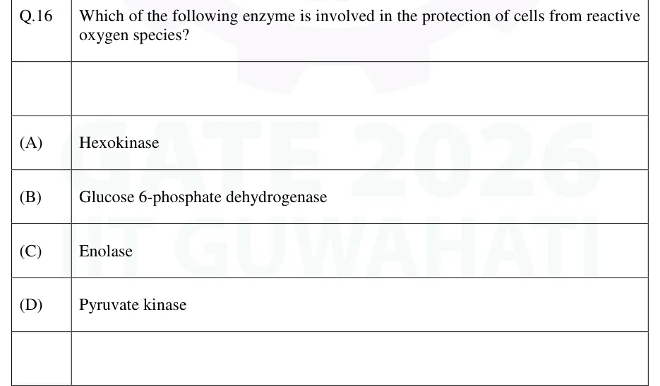
GATE 2026 BT Q.16
Which of the following enzyme is involved in the protection of cells from reactive oxygen species?
- (A)
Hexokinase
- (B)
Glucose 6-phosphate dehydrogenase
- (C)
Enolase
- (D)
Pyruvate kinase

GATE BT
Pathways, photosynthesis and respiration
Metabolism is how cells extract, store and spend energy. It covers the pathways that break down carbohydrates, lipids, amino acids and nucleotides and build them up again, and the light and carbon-fixation reactions that make sugar from CO2. These notes cover free energy and redox arithmetic, ATP and NADH yields (with the P/O convention stated), the regulated steps and their effectors, enzymes and cofactors, where in the cell each step happens, and how photosynthesis and respiration compare.
13 previous-year questions · 11 solved · 6 practice questions
A reaction proceeds spontaneously when its free energy change is negative. is the standard free energy change in the biochemical standard state: pH 7, 25 °C (298 K), 1 M of every other solute, and water taken as 1. The actual depends on the real concentrations, so a reaction with a positive can still run forward if its product is removed quickly. Standard free energy changes of consecutive reactions add.
FREE ENERGY UNDER CELLULAR CONDITIONS
R = 8.315 J/(mol·K); RT ≈ 2.48 kJ/mol at 25 °C. Q is the mass-action ratio [products]/[reactants]; at equilibrium Q = K'eq and ΔG = 0.
STANDARD FREE ENERGY AND EQUILIBRIUM CONSTANT
K'eq is the equilibrium constant at pH 7. Each tenfold change in K'eq changes ΔG°' by about 5.7 kJ/mol at 25 °C (this is RT ln 10).
FREE ENERGY OF A REDOX REACTION
n = electrons transferred per mole of the reaction as written, F = 96.5 kJ/(V·mol), ΔE°' = E°'(electron acceptor) minus E°'(electron donor), both at pH 7.
Coupled reactions add. An unfavourable reaction is driven by pairing it with ATP hydrolysis ( kJ/mol) through a shared intermediate. ATP is the energy currency because its hydrolysis is strongly favourable, not because it is a long-term store. In cells, ATP hydrolysis has of about −50 to −65 kJ/mol, because the ratio [ADP][Pi]/[ATP] is held far below its equilibrium value. A compound whose phosphate-group transfer is more favourable than ATP's (PEP, 1,3-bisphosphoglycerate, phosphocreatine) can make ATP from ADP directly. This is substrate-level phosphorylation.
Hydrolysis reaction | ΔG°' (kJ/mol, approximate) |
|---|---|
Phosphoenolpyruvate → pyruvate + Pi | −61.9 |
1,3-Bisphosphoglycerate → 3-phosphoglycerate + Pi | −49.3 |
ATP → AMP + PPi | −45.6 |
Phosphocreatine → creatine + Pi | −43.0 |
Acetyl-CoA → acetate + CoA (thioester) | −31.4 |
ATP → ADP + Pi | −30.5 |
Glucose-6-phosphate → glucose + Pi | −13.8 |
ADENYLATE ENERGY CHARGE
Ranges from 0 (all AMP) to 1 (all ATP); cells usually hold it at about 0.8–0.95. ATP-generating pathways are inhibited and ATP-using pathways stimulated as it rises. Adenylate kinase (2 ADP ⇌ ATP + AMP) makes [AMP] change far more, in proportion, than [ATP], which is why AMP is a sensitive low-energy signal.
Redox couple | E°' (V) |
|---|---|
NAD+/NADH | −0.32 |
NADP+/NADPH | −0.32 |
FAD/FADH2 (free) | about −0.22 |
Fumarate/succinate | +0.03 |
Ubiquinone/ubiquinol | +0.045 |
Cytochrome c (Fe3+/Fe2+) | +0.22 to +0.25 (sources differ) |
½ O2/H2O | +0.82 |
EXAM TIP
Electrons flow spontaneously from the couple with the more negative E°' to the one with the more positive E°'. If ΔE°' comes out negative in your calculation, you have the donor and acceptor swapped. Use n for the reaction as written: NADH gives 2 electrons.
Glycolysis occurs in the cytosol and converts one glucose into two pyruvate. Its investment phase spends two ATP and gives fructose-1,6-bisphosphate. Its payoff phase makes four ATP and two NADH per glucose. The net is 2 ATP, by substrate-level phosphorylation, and 2 NADH, from the oxidation of glyceraldehyde-3-phosphate. Overall: glucose + 2 NAD+ + 2 ADP + 2 Pi → 2 pyruvate + 2 NADH + 2 H+ + 2 ATP + 2 H2O.
Step | Enzyme | Note |
|---|---|---|
Glucose → G6P | Hexokinase (glucokinase in liver and pancreatic β cells) | Uses ATP; irreversible. Hexokinase has a low Km and is inhibited by G6P; glucokinase has a high Km and is not inhibited by G6P |
G6P ⇌ F6P | Phosphoglucose isomerase | Reversible |
F6P → F-1,6-BP | Phosphofructokinase-1 (PFK-1) | Uses ATP; irreversible; committed and main regulatory step |
F-1,6-BP ⇌ DHAP + G3P | Aldolase | Reversible; triose phosphate isomerase interconverts DHAP and G3P |
G3P → 1,3-BPG | Glyceraldehyde-3-phosphate dehydrogenase (GAPDH) | Makes NADH; uses Pi |
1,3-BPG → 3-PG | Phosphoglycerate kinase | Makes ATP (substrate level) |
3-PG ⇌ 2-PG ⇌ PEP | Phosphoglycerate mutase, enolase | Enolase is inhibited by fluoride |
PEP → pyruvate | Pyruvate kinase | Makes ATP (substrate level); irreversible |
Three steps are far from equilibrium and irreversible: hexokinase, PFK-1 and pyruvate kinase. These are the regulated steps. Hexokinase is inhibited by its product G6P. PFK-1 is inhibited by ATP, citrate and H+ (low pH), and activated by AMP, ADP and fructose-2,6-bisphosphate (F-2,6-BP). Pyruvate kinase is activated by F-1,6-BP (feed-forward). It is inhibited by ATP, acetyl-CoA and long-chain fatty acids, and, in liver, by alanine and by glucagon-triggered phosphorylation.
Fates of pyruvate: under aerobic conditions it enters mitochondria and is converted to acetyl-CoA by the pyruvate dehydrogenase complex (PDH). Without O2, cells must regenerate NAD+ so that glycolysis can continue. Animal cells reduce pyruvate to lactate (lactate dehydrogenase). Yeast decarboxylate it to acetaldehyde (pyruvate decarboxylase, which needs TPP) and reduce that to ethanol (alcohol dehydrogenase). Fermentation yields only the 2 ATP of glycolysis.
PDH sits in the mitochondrial matrix. Its components are E1 (pyruvate dehydrogenase, TPP), E2 (dihydrolipoyl transacetylase, lipoamide, CoA) and E3 (dihydrolipoyl dehydrogenase, FAD, NAD+). Reaction: pyruvate + CoA + NAD+ → acetyl-CoA + CO2 + NADH. It is irreversible, so acetyl-CoA cannot be turned back into pyruvate. PDH is inhibited by its products acetyl-CoA and NADH and by ATP. It is also inactivated by phosphorylation by PDH kinase, which acetyl-CoA, NADH and ATP activate and pyruvate and ADP inhibit. PDH phosphatase, activated by Ca2+ and insulin, reactivates it.
EXAM TIP
Arsenate replaces Pi at the GAPDH step. The product hydrolyses to 3-phosphoglycerate without making ATP, so glycolysis continues but its net ATP is zero. Arsenite is different: it binds the lipoamide of PDH and α-ketoglutarate dehydrogenase and inhibits both. Fluoride blocks enolase, and iodoacetate blocks GAPDH.
The citric acid cycle runs in the mitochondrial matrix (succinate dehydrogenase sits in the inner membrane). Acetyl-CoA (2 C) combines with oxaloacetate (4 C) to form citrate (6 C). Two carbons leave as CO2 in each turn, at isocitrate dehydrogenase and at α-ketoglutarate dehydrogenase. They are not the two carbons of the acetyl-CoA that entered in that turn; those leave in later turns. The cycle also supplies intermediates for biosynthesis. These are replenished by anaplerotic reactions: pyruvate carboxylase (animals), PEP carboxylase (plants and bacteria), malic enzyme and transamination.
Enzyme | Product made |
|---|---|
Citrate synthase | Citrate |
Aconitase | Isocitrate |
Isocitrate dehydrogenase | α-Ketoglutarate, CO2, NADH |
α-Ketoglutarate dehydrogenase (TPP, lipoamide, CoA, FAD, NAD+) | Succinyl-CoA, CO2, NADH |
Succinyl-CoA synthetase | Succinate, GTP (or ATP), by substrate-level phosphorylation |
Succinate dehydrogenase (Complex II) | Fumarate, FADH2 (FAD bound to the enzyme) |
Fumarase | Malate |
Malate dehydrogenase | Oxaloacetate, NADH |
Per acetyl-CoA, one turn gives 3 NADH, 1 FADH2, 1 GTP and 2 CO2. Regulation: citrate synthase is inhibited by NADH, succinyl-CoA and citrate (and ATP), and its rate also depends on oxaloacetate availability. Isocitrate dehydrogenase is inhibited by NADH and ATP and activated by ADP and Ca2+. α-Ketoglutarate dehydrogenase is inhibited by NADH and succinyl-CoA and activated by Ca2+.
Electron transport chain (inner mitochondrial membrane): NADH donates electrons to Complex I (NADH dehydrogenase), and FADH2 of succinate dehydrogenase enters at Complex II. Both pass electrons to the mobile carrier ubiquinone (Q). From Q they go to Complex III (cytochrome bc1), then the mobile protein cytochrome c on the intermembrane-space side, then Complex IV (cytochrome c oxidase; haem a, a3, CuA, CuB), which reduces O2 to H2O. Complexes I, III and IV move protons from the matrix to the intermembrane space; Complex II moves none. Complex III does this through the Q cycle. Complex IV pumps 2 H+ per 2 e− and takes 2 more H+ from the matrix to form water. Per pair of electrons this gives about 10 H+ from NADH and 6 from succinate. The resulting proton-motive force drives ATP synthase (F0F1) to make ATP (chemiosmosis).
Complex | Protons moved to the intermembrane space per 2 e− | Inhibitor |
|---|---|---|
I | 4 | Rotenone, amytal, piericidin A |
II | 0 | Malonate (competitive inhibitor of succinate dehydrogenase), carboxin |
III | 4 (Q cycle) | Antimycin A, myxothiazol |
IV | 2 (plus 2 taken up from the matrix to form water) | Cyanide, azide, CO |
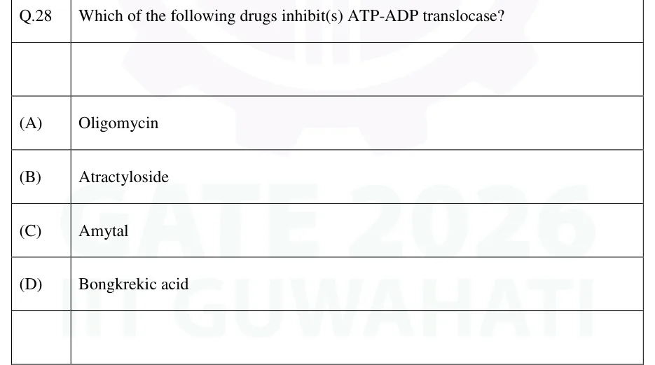
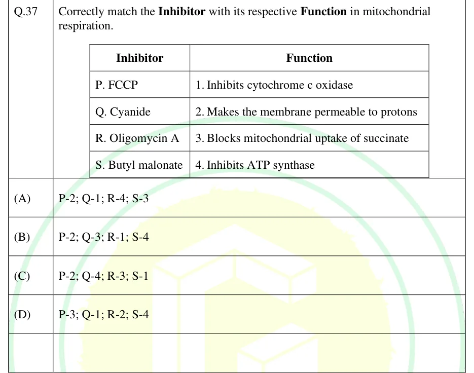
ATP synthase has a membrane F0 part (a c-ring that rotates as protons flow through) and a matrix F1 head (α3β3 with a central γ shaft). Rotation of γ drives each β site in turn through loose, tight and open states (binding-change mechanism), so one full rotation makes 3 ATP. The energy is needed mainly to release ATP from the enzyme. Oligomycin blocks F0. Atractyloside and bongkrekic acid block the ADP/ATP translocase. Uncouplers such as 2,4-dinitrophenol, and the protein thermogenin (UCP1) in brown fat, carry protons back across the membrane: oxygen consumption continues but ATP is not made, and the energy appears as heat. Respiratory control means that electron flow slows when ADP is scarce.
CONVENTION used here: P/O ratios of 2.5 ATP per NADH and 1.5 ATP per FADH2. These follow from about 10 and 6 protons moved per pair of electrons, and about 4 protons flowing back per ATP made and delivered to the cytosol: about 3 through ATP synthase (the exact number depends on the c-ring size) and 1 for the transport of Pi, ADP and ATP. Older texts use 3 and 2; when a question gives P/O ratios, use its values.
COMPLETE OXIDATION OF GLUCOSE (2.5 / 1.5 CONVENTION)
Terms: glycolysis ATP, cytosolic NADH (2.5 each via the malate-aspartate shuttle, 1.5 each via the glycerol-3-phosphate shuttle), PDH NADH, TCA GTP (counted as ATP), TCA NADH, TCA FADH2. Gives 32 with the malate-aspartate shuttle and 30 with the glycerol-3-phosphate shuttle.
Source | NADH | FADH2 | ATP/GTP directly |
|---|---|---|---|
Glycolysis (per glucose) | 2 (cytosolic) | 0 | 2 |
PDH (per glucose) | 2 | 0 | 0 |
TCA (per glucose, 2 turns) | 6 | 2 | 2 |
Total | 10 | 2 | 4 |
Cytosolic NADH cannot cross the inner membrane. The malate-aspartate shuttle (liver, heart, kidney) delivers its electrons as matrix NADH, worth 2.5 ATP. In the glycerol-3-phosphate shuttle (skeletal muscle, brain), mitochondrial glycerol-3-phosphate dehydrogenase, an FAD enzyme on the outer face of the inner membrane, passes the electrons straight to ubiquinone, bypassing Complex I. Each cytosolic NADH is then worth 1.5 rather than 2.5 ATP. With the older 3 and 2 ratios the totals are 38 and 36.
RESPIRATORY QUOTIENT
Carbohydrate 1.0 (C6H12O6 + 6 O2 → 6 CO2 + 6 H2O). Fat about 0.7 (palmitic acid: C16H32O2 + 23 O2 → 16 CO2 + 16 H2O, RQ = 16/23 ≈ 0.70). Protein about 0.8. Above 1 for oxygen-rich substrates such as organic acids (malate 4/3), when fat is being made from carbohydrate, or with fermentation.
EXAM TIP
Read what a question asks: ATP made, or net ATP. Then check whether it gives the P/O ratios and a shuttle. The same glucose is worth 30, 32, 36 or 38 ATP depending on these choices, and the answer is whichever the question's convention gives.
Gluconeogenesis makes glucose from pyruvate, lactate, glycerol and glucogenic amino acids, mainly in liver (and kidney). It reverses glycolysis except at the three irreversible steps, which are bypassed by four enzymes. Animals cannot make net glucose from even-chain fatty acids. PDH is irreversible, and each acetyl-CoA that enters the citric acid cycle adds two carbons while two leave as CO2, so no net oxaloacetate is gained. The glycerol of triacylglycerols and the propionyl-CoA from odd-chain fatty acids are glucogenic.
Glycolysis step bypassed | Gluconeogenic enzyme(s) | Location and note |
|---|---|---|
Pyruvate kinase | Pyruvate carboxylase, then PEP carboxykinase (PEPCK) | Carboxylase in mitochondria (biotin, ATP; activated by acetyl-CoA); PEPCK uses GTP (mitochondrial or cytosolic, depending on species) |
PFK-1 | Fructose-1,6-bisphosphatase (FBPase-1) | Cytosol; inhibited by AMP and F-2,6-BP |
Hexokinase | Glucose-6-phosphatase | Endoplasmic reticulum membrane of liver and kidney; absent from muscle |
Making one glucose from two pyruvate costs 4 ATP, 2 GTP (six high-energy phosphate bonds) and 2 NADH, while glycolysis makes only 2 ATP. The extra cost makes gluconeogenesis favourable under cellular conditions. If both pathways ran at full speed at once, they would form a futile (substrate) cycle that wastes ATP. This is prevented by reciprocal regulation of each bypass pair: AMP and F-2,6-BP activate PFK-1 and inhibit FBPase-1. The Cori cycle moves muscle lactate to liver for gluconeogenesis and returns glucose. The glucose-alanine cycle also carries amino nitrogen from muscle to liver.
Glyoxylate cycle: plants (in the glyoxysomes of germinating oilseeds), many bacteria and fungi can make net glucose from acetyl-CoA. Isocitrate lyase splits isocitrate into succinate and glyoxylate. Malate synthase joins glyoxylate with a second acetyl-CoA to give malate. The two decarboxylation steps of the citric acid cycle are skipped, so two acetyl-CoA yield one net four-carbon compound for gluconeogenesis. Animals lack both enzymes.
Glycogen breakdown: glycogen phosphorylase (cofactor pyridoxal phosphate) releases glucose-1-phosphate from non-reducing ends by phosphorolysis and stops four residues from a branch point. The debranching enzyme moves three of those residues to another chain (transferase activity) and releases the branch-point residue as free glucose (α-1,6-glucosidase activity). Phosphoglucomutase converts glucose-1-phosphate to G6P. Synthesis: UDP-glucose pyrophosphorylase makes UDP-glucose from glucose-1-phosphate and UTP. Glycogen synthase adds glucose in α-1,4 links to a primer (glycogenin starts a new particle), and the branching enzyme makes α-1,6 branches.
Hormonal control is reciprocal. Glucagon (liver) and epinephrine (liver and muscle) bind receptors and raise cAMP, which activates protein kinase A (PKA). PKA activates phosphorylase kinase, which converts phosphorylase b to the active a form; PKA also phosphorylates (inactivates) glycogen synthase. Insulin activates protein phosphatase 1, which reverses these. In muscle, Ca2+ activates phosphorylase kinase during contraction, and AMP activates phosphorylase b allosterically; ATP and G6P inhibit it. In liver, glucose inhibits phosphorylase a. G6P activates glycogen synthase. Likewise, glucagon through PKA phosphorylates the liver's bifunctional PFK-2/FBPase-2, lowering F-2,6-BP, so glycolysis slows and gluconeogenesis rises. Insulin raises F-2,6-BP.
The pentose phosphate pathway (cytosol) has two parts. The oxidative part: glucose-6-phosphate dehydrogenase (G6PD; the committed step, controlled by the NADP+/NADPH ratio and inhibited by NADPH), lactonase and 6-phosphogluconate dehydrogenase. Together they give ribulose-5-phosphate, 2 NADPH and 1 CO2 per glucose-6-phosphate. The non-oxidative part interconverts sugars from 3 to 7 carbons and links back to glycolysis: transketolase (needs TPP) moves 2-carbon units, and transaldolase moves 3-carbon units. The pathway supplies NADPH for fatty acid and steroid synthesis and for glutathione reduction, and ribose-5-phosphate for nucleotides. It is active in liver, adipose tissue, adrenal cortex and red cells. No ATP is made or used directly.
EXAM TIP
Glucose-6-phosphate dehydrogenase deficiency leaves red cells short of NADPH and so of reduced glutathione, making them fragile under oxidative stress.
Fatty acid β-oxidation takes place in the mitochondrial matrix. First the fatty acid is activated outside the matrix: acyl-CoA synthetase (outer mitochondrial membrane) converts it to acyl-CoA using ATP, which becomes AMP + PPi, equal to 2 ATP equivalents. Long-chain acyl groups enter on carnitine: CPT-I on the outer membrane (inhibited by malonyl-CoA), the carnitine-acylcarnitine translocase, then CPT-II on the inner membrane. Each round of four reactions releases one acetyl-CoA and shortens the chain by two carbons: acyl-CoA dehydrogenase (FADH2), enoyl-CoA hydratase, 3-hydroxyacyl-CoA dehydrogenase (NADH), thiolase.
COMPLETE OXIDATION OF AN EVEN-CHAIN SATURATED FATTY ACID WITH C CARBONS (C ≥ 4)
Uses 2.5 per NADH and 1.5 per FADH2, so 4 per β-oxidation round and 10 per acetyl-CoA, and 2 spent on activation. For palmitate (C = 16): 7 rounds give 28, 8 acetyl-CoA give 80, gross 108, net 106. Stearate (C = 18) gives 120. With the older ratios (3 and 2) palmitate gives 129.
An odd-chain fatty acid ends in propionyl-CoA. Propionyl-CoA carboxylase (biotin) makes D-methylmalonyl-CoA, an epimerase gives the L form, and methylmalonyl-CoA mutase (deoxyadenosylcobalamin, a vitamin B12 form) makes succinyl-CoA, which is glucogenic. A cis double bond at an odd-numbered carbon (oleate's Δ9) needs enoyl-CoA isomerase and skips one acyl-CoA dehydrogenase step. One fewer FADH2 is made, so the yield is 1.5 ATP lower in this convention (oleate 118.5). A double bond at an even-numbered carbon also needs 2,4-dienoyl-CoA reductase, which uses NADPH.
Ketone bodies: in prolonged fasting, starvation or uncontrolled diabetes, liver mitochondria convert excess acetyl-CoA into acetoacetate through HMG-CoA (mitochondrial HMG-CoA synthase). Acetoacetate is reduced to β-hydroxybutyrate; acetone forms from acetoacetate as a minor by-product. The liver releases ketone bodies but cannot use them, because it lacks succinyl-CoA:3-ketoacid CoA transferase (thiophorase). Heart and skeletal muscle oxidise them, and so does the brain in prolonged fasting.
Fatty acid synthesis is cytosolic and is not the reverse of β-oxidation. Acetyl-CoA leaves the mitochondrion as citrate, and citrate lyase regenerates it in the cytosol. Acetyl-CoA carboxylase (ACC; biotin, ATP) makes malonyl-CoA. This is the committed step, activated by citrate and insulin and inhibited by palmitoyl-CoA and by phosphorylation (AMPK; PKA after glucagon or epinephrine). Fatty acid synthase adds two-carbon units from malonyl-CoA, using NADPH from the pentose phosphate pathway and malic enzyme, and releases palmitate. Palmitate needs 8 acetyl-CoA, 7 ATP (for the 7 malonyl-CoA) and 14 NADPH.
Cholesterol is made in the cytosol and endoplasmic reticulum from acetyl-CoA. HMG-CoA comes from a cytosolic HMG-CoA synthase (the ketogenic one is mitochondrial). HMG-CoA reductase (endoplasmic reticulum membrane, uses 2 NADPH) then converts HMG-CoA to mevalonate. This is the committed and main regulated step. The enzyme is inhibited by cholesterol (feedback, including lower synthesis of the enzyme), by phosphorylation (AMPK) and by statins, which are competitive inhibitors.
Feature | β-oxidation | Fatty acid synthesis |
|---|---|---|
Location | Mitochondrial matrix | Cytosol |
Carrier of acyl group | CoA | Acyl carrier protein domain of fatty acid synthase |
Redox cofactor | NAD+ and FAD accept electrons | NADPH donates electrons |
3-Hydroxyacyl intermediate | L isomer | D isomer |
Two-carbon unit | Acetyl-CoA released | Malonyl-CoA donates |
Regulated by | Malonyl-CoA blocks CPT-I | Acetyl-CoA carboxylase |
Amino acid catabolism begins with removal of the α-amino group. Transaminases (pyridoxal phosphate) move it to α-ketoglutarate, giving glutamate. Glutamate dehydrogenase (NAD+ or NADP+) then releases free NH4+ by oxidative deamination in mitochondria. The carbon skeletons enter metabolism as pyruvate, acetyl-CoA or acetoacetyl-CoA, α-ketoglutarate, succinyl-CoA, fumarate or oxaloacetate. Leucine and lysine are purely ketogenic. Isoleucine, phenylalanine, tyrosine and tryptophan are both ketogenic and glucogenic, and some texts also class threonine as both. The rest are glucogenic.
Urea cycle, in order
Carbamoyl phosphate synthetase I (mitochondria, needs 2 ATP, activated by N-acetylglutamate) makes carbamoyl phosphate from NH4+ and HCO3−.
Ornithine transcarbamoylase (mitochondria) joins carbamoyl phosphate to ornithine to make citrulline.
Citrulline moves to the cytosol; argininosuccinate synthetase (ATP → AMP + PPi) adds aspartate to give argininosuccinate.
Argininosuccinate lyase releases fumarate and arginine.
Arginase hydrolyses arginine to urea and regenerates ornithine, which returns to the mitochondria.
The cycle costs four high-energy phosphate bonds per urea (3 ATP, one of them becoming AMP + PPi). Of urea's two nitrogens, one comes from NH4+ (via carbamoyl phosphate) and one from aspartate. The fumarate released is converted to malate and oxaloacetate, which can be transaminated back to aspartate. This links the urea cycle to the citric acid cycle.
Purines: de novo synthesis builds the ring step by step on ribose-5-phosphate, starting from PRPP, to make IMP. The committed enzyme, glutamine-PRPP amidotransferase, is activated by PRPP and inhibited by IMP, AMP and GMP. IMP becomes AMP (needs GTP) or GMP (needs ATP). Salvage by HGPRT recycles hypoxanthine and guanine, and its absence causes Lesch-Nyhan syndrome. Purines are degraded to uric acid by xanthine oxidase, which allopurinol inhibits.
Pyrimidines: the ring is built first, from carbamoyl phosphate (CPS II, cytosol) and aspartate (aspartate transcarbamoylase, ATCase), giving orotate. Orotate is then attached to PRPP and decarboxylated to UMP; CTP comes from UTP (CTP synthetase, glutamine). In E. coli, ATCase is the regulated step: CTP inhibits it and ATP activates it. In animals, CPS II is regulated: UTP inhibits it and PRPP and ATP activate it. Ribonucleotide reductase converts NDPs to dNDPs, and dATP inhibits its overall activity. Thymidylate synthase makes dTMP from dUMP using N5,N10-methylene tetrahydrofolate. It is blocked by FdUMP, formed from 5-fluorouracil. Dihydrofolate reductase is blocked by methotrexate. Pyrimidines are degraded to CO2, NH4+ and β-alanine or β-aminoisobutyrate.
Where the ring atoms come from
Purine: N1 from aspartate; C2 and C8 from N10-formyl tetrahydrofolate; N3 and N9 from the amide N of glutamine; C4, C5 and N7 from glycine; C6 from CO2 (HCO3−).
Pyrimidine: N1, C4, C5 and C6 from aspartate; C2 from HCO3− and N3 from glutamine, both via carbamoyl phosphate.
Pathway | Main regulated enzyme | Activated by | Inhibited by |
|---|---|---|---|
Glycolysis | PFK-1 | AMP, ADP, F-2,6-BP | ATP, citrate, H+ |
Pyruvate → acetyl-CoA | PDH complex | Dephosphorylation (PDH phosphatase; Ca2+, insulin) | Acetyl-CoA, NADH, ATP; phosphorylation (PDH kinase) |
Citric acid cycle | Isocitrate dehydrogenase (also citrate synthase, α-ketoglutarate dehydrogenase) | ADP, Ca2+ | NADH, ATP |
Gluconeogenesis | Pyruvate carboxylase; FBPase-1 | Acetyl-CoA (pyruvate carboxylase) | AMP, F-2,6-BP (FBPase-1) |
Glycogen breakdown | Glycogen phosphorylase | Phosphorylation (glucagon, epinephrine); AMP and, via phosphorylase kinase, Ca2+ (muscle) | Glucose (liver); ATP, G6P (muscle) |
Glycogen synthesis | Glycogen synthase | Dephosphorylation (insulin); G6P | Phosphorylation (glucagon, epinephrine) |
Pentose phosphate pathway | Glucose-6-phosphate dehydrogenase | NADP+ | NADPH |
Fatty acid synthesis | Acetyl-CoA carboxylase | Citrate, insulin | Palmitoyl-CoA; phosphorylation (AMPK, PKA) |
β-oxidation | CPT-I (carnitine entry) | — | Malonyl-CoA |
Cholesterol synthesis | HMG-CoA reductase | — | Cholesterol, statins, phosphorylation (AMPK) |
Urea cycle | Carbamoyl phosphate synthetase I | N-acetylglutamate | — |
Purine synthesis | Glutamine-PRPP amidotransferase | PRPP | IMP, AMP, GMP |
Pyrimidine synthesis | ATCase (E. coli); CPS II (animals) | ATP (ATCase); PRPP, ATP (CPS II) | CTP (ATCase); UTP (CPS II) |
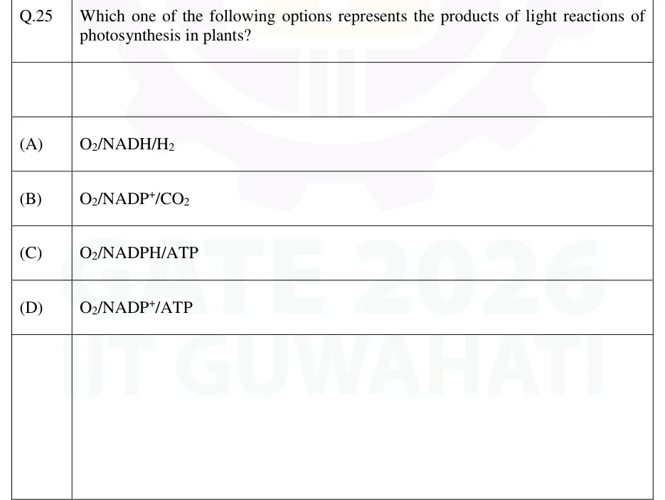
In plants, photosynthesis occurs in chloroplasts. The light reactions in the thylakoid membrane turn light into ATP and NADPH and release O2. The Calvin cycle in the stroma uses that ATP and NADPH to fix CO2. Overall: 6 CO2 + 12 H2O → C6H12O6 + 6 O2 + 6 H2O. Labelling with H2(18)O shows that the O2 released comes from water, not from CO2.
Z-scheme (non-cyclic electron flow): photosystem II (reaction centre P680) is excited. Its water-oxidising complex (a Mn4CaO5 cluster) oxidises , releasing the protons into the lumen. Electrons go through pheophytin and the quinones QA and QB to the plastoquinone pool, then to the cytochrome b6f complex, which moves protons into the lumen by a Q cycle. Plastocyanin carries them on to photosystem I (P700). A second light excitation drives them through ferredoxin to ferredoxin-NADP+ reductase, which makes NADPH. Protons accumulate in the lumen (about pH 5, against about pH 8 in the stroma in light). ATP synthase (CF0CF1) lets them flow back to the stroma and makes ATP (photophosphorylation).
About 8 photons are absorbed per O2 released, 4 by each photosystem, because each of the 4 electrons taken from 2 H2O is excited twice. Non-cyclic flow per O2 gives 2 NADPH and about 3 ATP (the ATP figure depends on the proton stoichiometry assumed). Light beyond about 680 nm drives photosystem I but hardly photosystem II, so the yield falls in far-red light (red drop). Adding shorter-wavelength light raises the rate above the sum of the two beams given separately (enhancement effect). This is evidence that the two photosystems work in series.
Cyclic electron flow uses only photosystem I: electrons return from ferredoxin to cytochrome b6f and back to P700. It makes ATP only, with no NADPH and no O2. It tops up the ATP supply, because the Calvin cycle needs ATP and NADPH in the ratio 3 : 2.
Calvin cycle phases
Carboxylation: RuBisCO attaches CO2 to ribulose-1,5-bisphosphate (RuBP, 5 C), giving two molecules of 3-phosphoglycerate (3-PGA).
Reduction: 3-PGA is phosphorylated by ATP (phosphoglycerate kinase) and reduced by NADPH (NADP-dependent GAPDH) to glyceraldehyde-3-phosphate (G3P).
Regeneration: five of every six G3P are rearranged to regenerate three RuBP, using ATP for the final phosphorylation (phosphoribulokinase); the sixth G3P is the net product.
CALVIN CYCLE STOICHIOMETRY
Per CO2 fixed: 3 ATP and 2 NADPH. Three CO2 give one net G3P using 9 ATP and 6 NADPH.
Light switches on the Calvin cycle enzymes. Stromal pH rises to about 8 and stromal Mg2+ rises. Ferredoxin-thioredoxin reduction activates FBPase, sedoheptulose-1,7-bisphosphatase, phosphoribulokinase and GAPDH. RuBisCO must be carbamylated: a CO2 molecule (separate from the substrate CO2) reacts with an active-site lysine, and Mg2+ then binds. RuBisCO activase removes inhibitory sugar phosphates from the active site.
Photorespiration: RuBisCO also accepts O2. RuBP + O2 gives one 3-PGA and one 2-phosphoglycolate, which is recycled through chloroplast, peroxisome and mitochondria (the C2 cycle). The cycle releases CO2 and NH3 when two glycine become serine in the mitochondria. For every two 2-phosphoglycolate (4 C), one carbon is lost as CO2 and three return to the Calvin cycle as 3-PGA. Photorespiration uses ATP and reducing power without making sugar. It rises with temperature and with the O2 to CO2 ratio.
Feature | C3 | C4 | CAM |
|---|---|---|---|
First product of fixation | 3-PGA (RuBisCO) | Oxaloacetate (PEP carboxylase in mesophyll) | Oxaloacetate, then malate (PEP carboxylase at night) |
Where the Calvin cycle runs | Mesophyll | Bundle sheath | Same cell, by day |
How CO2 is separated | None | Spatial: malate or aspartate carries CO2 to the bundle sheath (Kranz anatomy) | Temporal: malate stored in the vacuole at night, decarboxylated by day |
Stomata open | Day | Day | Night |
ATP per CO2 fixed | 3 | 5 (extra 2 for PEP regeneration by pyruvate phosphate dikinase) | More than 3 (PEP is regenerated from stored carbohydrate each night) |
Photorespiration | Significant, rising with temperature | Very low | Very low by day |
CO2 compensation point | High | Very low | Low |
Examples | Wheat, rice | Maize, sugarcane | Cactus, pineapple |
EXAM TIP
C4 and CAM plants avoid most photorespiration because CO2 is concentrated around RuBisCO, in the bundle sheath or behind closed stomata by day. This suppresses RuBisCO's oxygenase activity. The first carboxylase, PEP carboxylase, fixes bicarbonate (HCO3−), not CO2 itself, and does not react with O2.
Property | Respiration (mitochondria) | Photosynthesis (chloroplast) |
|---|---|---|
Electron carrier | NADH, FADH2 | NADPH |
Electron donor and final acceptor | Fuel (via NADH, FADH2) to O2 (becomes H2O) | H2O (becomes O2) to NADP+ |
Protons accumulate in | Intermembrane space | Thylakoid lumen |
Proton-motive force mainly as | Membrane potential | pH gradient |
ATP synthase F1 head faces | Matrix | Stroma |
Energy source | Oxidation of fuels | Light |
PREVIOUS-YEAR QUESTIONS
Try each question first, then open the answer. We placed each question under a topic ourselves; the exam paper does not label topics.
See these papers in full: GATE BT 2026 · GATE BT 2025 · GATE BT 2024 · GATE BT 2022 · GATE BT 2020
Which of the following enzyme is involved in the protection of cells from reactive oxygen species?
Hexokinase
Glucose 6-phosphate dehydrogenase
Enolase
Pyruvate kinase
Which one of the following options represents the products of light reactions of photosynthesis in plants?
O2/NADH/H2
O2/NADP+/CO2
O2/NADPH/ATP
O2/NADP+/ATP
Which of the following drugs inhibit(s) ATP-ADP translocase?
Oligomycin
Atractyloside
Amytal
Bongkrekic acid
Correctly match the Inhibitor with its respective Function in mitochondrial respiration.
Inhibitor | Function
P. FCCP | 1. Inhibits cytochrome c oxidase
Q. Cyanide | 2. Makes the membrane permeable to protons
R. Oligomycin A | 3. Blocks mitochondrial uptake of succinate
S. Butyl malonate | 4. Inhibits ATP synthase
P-2; Q-1; R-4; S-3
P-2; Q-3; R-1; S-4
P-2; Q-4; R-3; S-1
P-3; Q-1; R-2; S-4
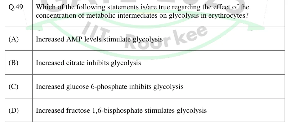
Which of the following statements is/are true regarding the effect of the concentration of metabolic intermediates on glycolysis in erythrocytes?
Increased AMP levels stimulate glycolysis
Increased citrate inhibits glycolysis
Increased glucose 6-phosphate inhibits glycolysis
Increased fructose 1,6-bisphosphate stimulates glycolysis
Official answer: A, B, DSource: Official answer key, GATE 2025 BT (IIT Roorkee)
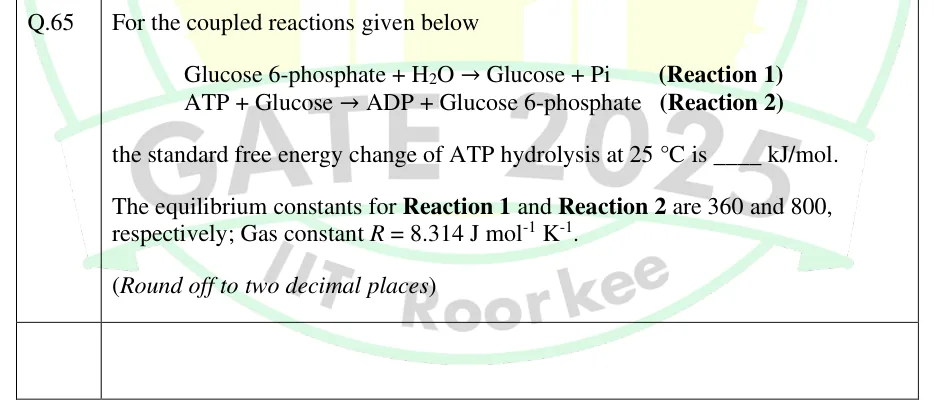
For the coupled reactions given below
Glucose 6-phosphate + H2O → Glucose + Pi (Reaction 1)
ATP + Glucose → ADP + Glucose 6-phosphate (Reaction 2)
the standard free energy change of ATP hydrolysis at 25 °C is ____ kJ/mol.
The equilibrium constants for Reaction 1 and Reaction 2 are 360 and 800, respectively; Gas constant R = 8.314 J mol^-1 K^-1.
(Round off to two decimal places)
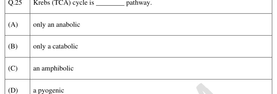
Krebs (TCA) cycle is ________ pathway.
only an anabolic
only a catabolic
an amphibolic
a pyogenic
The free energy change of ATP hydrolysis at 25 °C is -32.2 kJ mol^-1. The free energy change for hydrolysis of α-glycerophosphate to glycerol is -8.2 kJ mol^-1 at 25 °C. Using the above information, the free energy change for the formation of α-glycerophosphate from glycerol and ATP is ________ kJ mol^-1 (Answer in integer).
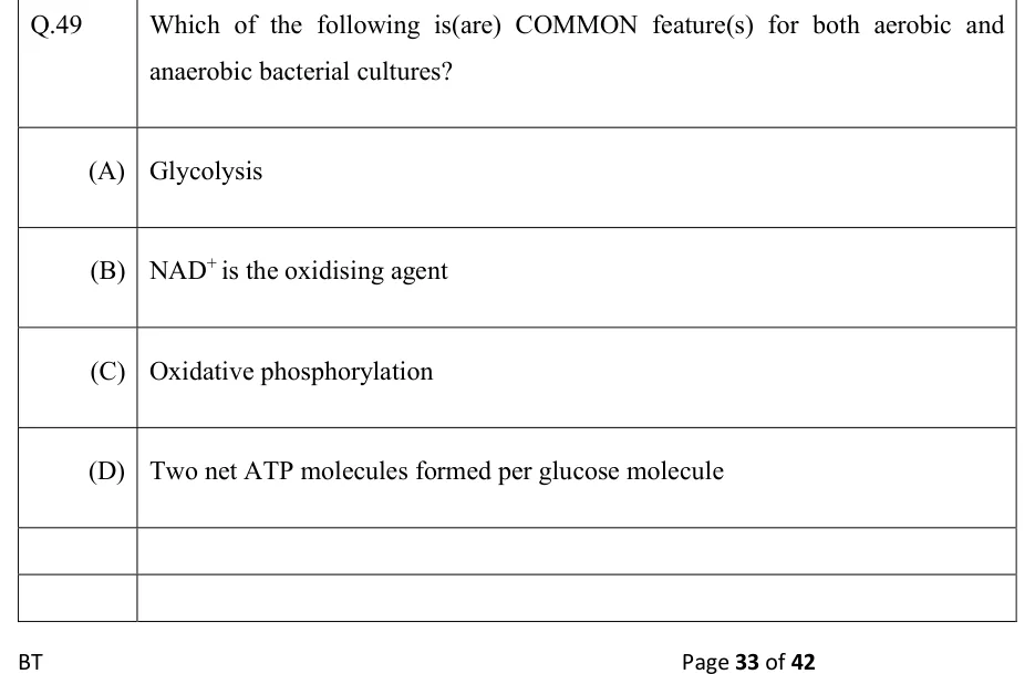
Which of the following is(are) COMMON feature(s) for both aerobic and anaerobic bacterial cultures?
Glycolysis
is the oxidising agent
Oxidative phosphorylation
Two net ATP molecules formed per glucose molecule
Official answer: A, BSource: Official answer key, GATE 2022 BT (IIT Kharagpur)
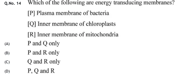
Which of the following are energy transducing membranes?
[P] Plasma membrane of bacteria
[Q] Inner membrane of chloroplasts
[R] Inner membrane of mitochondria
P and Q only
P and R only
Q and R only
P, Q and R
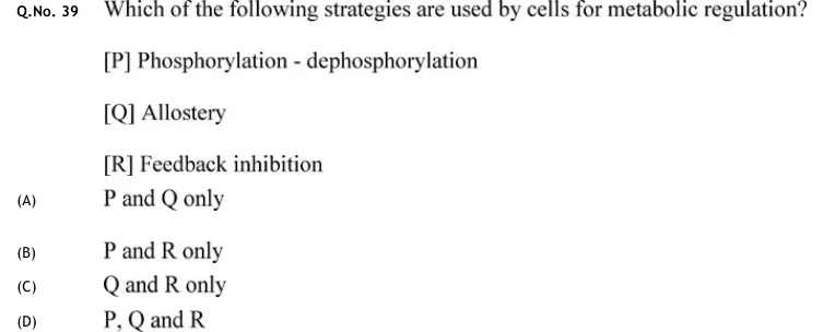
Which of the following strategies are used by cells for metabolic regulation?
[P] Phosphorylation - dephosphorylation
[Q] Allostery
[R] Feedback inhibition
P and Q only
P and R only
Q and R only
P, Q and R
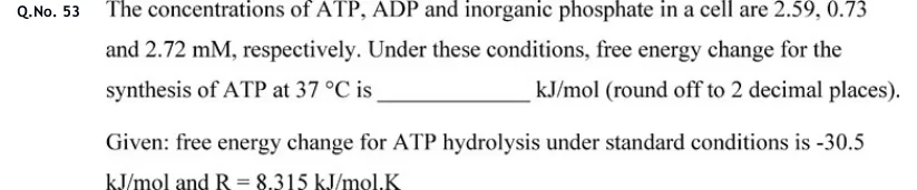
The concentrations of ATP, ADP and inorganic phosphate in a cell are 2.59, 0.73 and 2.72 mM, respectively. Under these conditions, free energy change for the synthesis of ATP at 37 °C is ______ kJ/mol (round off to 2 decimal places). Given: free energy change for ATP hydrolysis under standard conditions is -30.5 kJ/mol and R = 8.315 kJ/mol.K
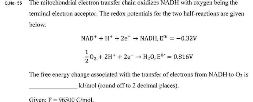
The mitochondrial electron transfer chain oxidizes NADH with oxygen being the terminal electron acceptor. The redox potentials for the two half-reactions are given below:
,
,
The free energy change associated with the transfer of electrons from NADH to is ______ kJ/mol (round off to 2 decimal places).
Given: .
PRACTICE
Using P/O ratios of 2.5 ATP per NADH and 1.5 ATP per FADH2, and counting the 2 ATP equivalents spent on activation, how many ATP are made net by complete oxidation of one molecule of palmitic acid (C16, saturated) to CO2 and H2O?
Answer:
Palmitate (16 C) undergoes 7 rounds of β-oxidation, giving 7 FADH2, 7 NADH and 8 acetyl-CoA.
7 FADH2 × 1.5 = 10.5 ATP; 7 NADH × 2.5 = 17.5 ATP.
8 acetyl-CoA × 10 ATP (3 NADH, 1 FADH2, 1 GTP per turn) = 80 ATP.
Gross = 10.5 + 17.5 + 80 = 108; subtract 2 for activation: net = 106.
Electrons pass from NADH (E°' = −0.32 V) to O2 (E°' = +0.82 V for the ½ O2/H2O couple) through the respiratory chain. Taking F = 96.5 kJ/(V·mol), find the magnitude of ΔG°' for oxidation of one mole of NADH by ½ O2, in kJ/mol (to the nearest whole number).
Answer: (accepted range )
ΔE°' = E°'(acceptor) − E°'(donor) = 0.82 − (−0.32) = 1.14 V; n = 2.
ΔG°' = −nFΔE°' = −2 × 96.5 × 1.14 ≈ −220 kJ/mol.
The magnitude is 220 kJ/mol.
In a C4 plant such as maize, inorganic carbon from atmospheric CO2 is first fixed (as HCO3−) in mesophyll cells. Which pair gives the enzyme and the first stable product?
RuBisCO; 3-phosphoglycerate
PEP carboxylase; oxaloacetate
PEP carboxylase; 3-phosphoglycerate
RuBisCO; oxaloacetate
Answer: (B)
In C4 plants, bicarbonate is added to phosphoenolpyruvate by PEP carboxylase in mesophyll cells.
The product is the four-carbon oxaloacetate, later converted to malate or aspartate.
RuBisCO acts later, in bundle-sheath cells, which excludes A and D; PEP carboxylase does not make 3-phosphoglycerate, which excludes C.
In a C3 plant, the Calvin cycle fixes 6 molecules of CO2 into one hexose. Counting only the cycle itself, what is the number of ATP molecules consumed plus the number of NADPH molecules consumed?
Answer:
Each CO2 fixed needs 3 ATP and 2 NADPH.
For 6 CO2: 18 ATP and 12 NADPH.
Sum = 18 + 12 = 30.
Calculate the respiratory quotient (moles of CO2 released per mole of O2 consumed) for the complete oxidation of palmitic acid, C16H32O2, to CO2 and H2O. Give the answer to two decimal places.
Answer: (accepted range )
Balance the equation: C16H32O2 + 23 O2 → 16 CO2 + 16 H2O.
Check oxygen: left 2 + 46 = 48; right 32 + 16 = 48. Hydrogen: 32 on each side.
RQ = 16/23 ≈ 0.696, which is 0.70 to two decimal places.
Which of the following reactions in mammalian liver are bypass steps unique to gluconeogenesis, that is, not simple reversals of glycolytic reactions?
Pyruvate → oxaloacetate, catalysed by pyruvate carboxylase
1,3-Bisphosphoglycerate → 3-phosphoglycerate, catalysed by phosphoglycerate kinase
Fructose-1,6-bisphosphate → fructose-6-phosphate, catalysed by fructose-1,6-bisphosphatase
Glucose → glucose-6-phosphate, catalysed by hexokinase
Answer: (A), (C)
Pyruvate carboxylase (with PEP carboxykinase) bypasses pyruvate kinase, so A is correct.
Phosphoglycerate kinase works reversibly in both pathways, so B is not a bypass.
Fructose-1,6-bisphosphatase bypasses PFK-1, so C is correct.
Hexokinase is a glycolytic enzyme; the gluconeogenic bypass is glucose-6-phosphatase, so D is wrong.
OFFICIAL GATE 2027 SYLLABUS WORDING
Copied from the GATE 2027 syllabus; see all 2027 syllabi.
GATE BT
Basic concepts and regulation of metabolism of carbohydrates, lipids, amino acids and nucleic acids; Metabolism: Regulation of metabolism of carbohydrates, lipids, amino acids, and nucleic acids; Detailed pathways like glycolysis, citric acid cycle, and fatty acid oxidation; Photosynthesis, respiration and electron transport chain
Preparing in Kolkata? IAE Academy's course for GATE BT.