GATE 2026 BT Q.48
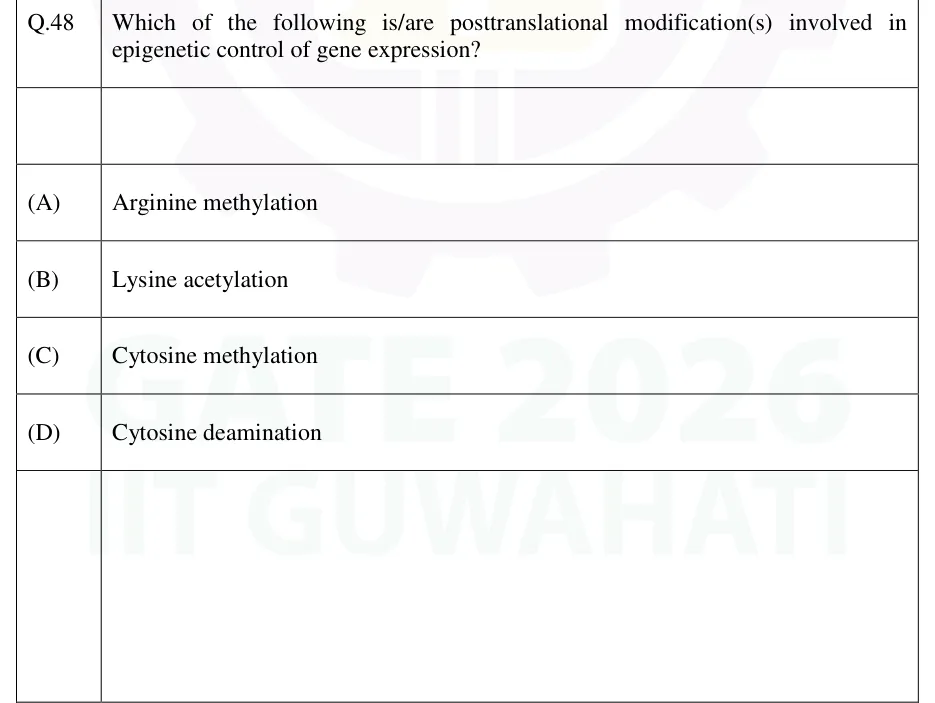
Which of the following is/are posttranslational modification(s) involved in epigenetic control of gene expression?
- (A)
Arginine methylation
- (B)
Lysine acetylation
- (C)
Cytosine methylation
- (D)
Cytosine deamination

GATE BT
Cell communication and signal transduction cover how cells send and receive signals (endocrine, paracrine, synaptic, autocrine, contact-dependent, gap junctions and plasmodesmata, bacterial quorum sensing). They also cover how the main receptor classes (GPCRs, receptor tyrosine kinases, cytokine receptors, receptor serine/threonine kinases, ligand-gated ion channels and nuclear receptors) turn a ligand into an intracellular response through G proteins, second messengers and kinase cascades, and how those signals are switched off. The topic includes the quantitative description of receptor-ligand binding (, fractional occupancy, the Scatchard and Hill equations), what makes a gene's effect non-cell-autonomous, and the post-translational modifications that turn signalling proteins on and off.
7 previous-year questions · 6 solved · 6 practice questions
Cells almost never act alone. They constantly send and receive chemical signals that coordinate growth, metabolism, development and stress responses. This topic covers how a signal (a ligand) is detected by a receptor, turned into an intracellular message by G proteins, second messengers and kinase cascades, and eventually switched off. It also covers how a gene's effect can appear in cells other than the ones that express it (non-cell-autonomous signalling), and how post-translational modifications switch the proteins involved on and off, move them or destroy them.
Modes of signalling, by distance travelled and route
Endocrine: a hormone is secreted into the bloodstream and acts on distant target cells that carry the matching receptor (e.g. insulin from the pancreas acting on liver, muscle and fat).
Paracrine: a secreted signal diffuses through the local extracellular fluid and acts only on nearby cells, without entering the circulation (e.g. growth factors acting on neighbouring cells during development).
Synaptic (neuronal): a neuron carries the signal over a long distance as an electrical impulse along its axon. At the synapse it releases a neurotransmitter that acts on one specific target cell a very short distance away.
Autocrine: a cell secretes a signal that binds receptors on itself (or on cells of the same type), a self-stimulating loop seen, for example, in some immune cells and cancer cells.
Contact-dependent (juxtacrine): the ligand stays anchored in the membrane of the signalling cell and binds a receptor on an adjacent cell, so the two cells must touch. For example, Delta on the signalling cell binds Notch on the receiving cell. No secretion or diffusion is involved.
Gap junctions (animals) and plasmodesmata (plants): direct cytoplasm-to-cytoplasm channels between adjacent cells. A gap-junction channel is made of two connexons (one from each cell), each built from six connexin subunits. It lets ions and small water-soluble molecules through (roughly 1 kDa or less, e.g. , cAMP, ) but not proteins or nucleic acids. Plasmodesmata can also let some proteins and RNAs pass.
Quorum sensing (bacteria): cells secrete small autoinducer molecules whose concentration rises with population density. Above a threshold concentration, the population switches on genes together (e.g. bioluminescence, biofilm formation, virulence). Gram-negative bacteria commonly use acyl-homoserine lactones (AHLs); Gram-positive bacteria commonly use modified oligopeptides.
EXAM TIP
Classify by where the ligand goes. Carried in the blood to distant cells: endocrine. Secreted and acting on nearby other cells: paracrine. Acting back on the cell that released it: autocrine. Held in the signalling cell's membrane, so the cells must touch: contact-dependent (juxtacrine). Passing straight from one cytoplasm to the next: gap junction / plasmodesma.
Every receptor turns an extracellular (or lipophilic) signal into a change in cell behaviour, but the classes differ in mechanism, location and typical response speed. Cell-surface receptors bind water-soluble ligands that cannot cross the membrane. Intracellular (nuclear) receptors bind small lipophilic ligands that reach the inside of the cell.
Receptor classes compared
Receptor class | Representative ligands | Mechanism | Location | Typical response speed |
|---|---|---|---|---|
G-protein-coupled receptor (GPCR; 7-transmembrane) | Adrenaline (epinephrine), glucagon, many neurotransmitters, odorants, light (rhodopsin) | The ligand-bound receptor acts as a guanine-nucleotide exchange factor (GEF) for a heterotrimeric G protein: releases GDP and binds GTP. The receptor has no enzymatic domain of its own. | Plasma membrane | Seconds to minutes |
Receptor tyrosine kinase (RTK) | Insulin, EGF, PDGF, NGF | Ligand binding brings two kinase domains together. Most RTKs dimerize on ligand binding; the insulin receptor is already a disulfide-linked dimer and is activated by a conformational change. The kinase domains cross-phosphorylate each other's tyrosines (trans-autophosphorylation), and the phosphotyrosines become docking sites for SH2/PTB-domain proteins such as the adaptor Grb2. | Plasma membrane | Minutes (direct kinase effects) to hours (effects through gene expression) |
Cytokine receptor (tyrosine-kinase-associated) | Interferons, interleukins, growth hormone, erythropoietin | No kinase domain of its own. Ligand binding brings together non-covalently associated JAK tyrosine kinases, which phosphorylate each other, the receptor and STAT proteins. | Plasma membrane | Minutes (STAT-driven transcription) |
Receptor serine/threonine kinase | TGF-β superfamily (including BMPs) | The ligand brings type II and type I receptors together. The type II receptor phosphorylates the type I receptor, which then phosphorylates SMAD transcription regulators. | Plasma membrane | Minutes to hours |
Ligand-gated ion channel | Acetylcholine (nicotinic receptor), GABA ( receptor), glycine | Ligand binding directly opens an ion-conducting pore in the receptor itself. | Plasma membrane | Milliseconds |
Nuclear (intracellular) receptor | Steroid hormones (cortisol, oestrogen, testosterone), thyroid hormone, retinoic acid, vitamin D (calcitriol) | The ligand reaches the receptor inside the cell: steroids diffuse across the membrane, and thyroid hormone enters through membrane transporters. The ligand-bound receptor binds hormone response elements in DNA and acts directly as a ligand-activated transcription factor. | Cytoplasm (e.g. glucocorticoid receptor, held by Hsp90 until ligand binds) or nucleus (e.g. thyroid hormone receptor) | Typically tens of minutes to hours (new transcription and translation needed) |
EXAM TIP
An RTK carries its own tyrosine kinase domain. A cytokine receptor has none and relies on associated JAKs. A GPCR has no enzymatic activity at all: it activates a separate G protein. Only the ligand-gated ion channel is itself the effector.
Binding at equilibrium is described by the dissociation constant . Convention used below: is free receptor, is free ligand, is the receptor–ligand complex, and . All concentrations are free concentrations at equilibrium. When a question gives only the ligand added, it is assumed that binding does not measurably deplete the free ligand.
EQUILIBRIUM DISSOCIATION CONSTANT
For a simple 1:1 interaction. has units of concentration ( in , in ). A smaller means tighter (higher-affinity) binding. The association constant is .
FRACTIONAL RECEPTOR OCCUPANCY (SINGLE SITE, NON-COOPERATIVE)
At , . Rearranged, , so moving from 10% to 90% occupancy needs an 81-fold rise in (from to ). Valid only for one independent binding site per receptor.
SCATCHARD EQUATION
= bound ligand (), = free ligand, = total receptor concentration. For a single class of independent sites, plotting against gives a straight line with slope and x-intercept . A curved Scatchard plot indicates cooperativity or more than one class of binding site.
HILL EQUATION (COOPERATIVE BINDING)
Here has units of concentration and equals , where is the ligand concentration that gives half saturation. is the Hill coefficient: means positive cooperativity, means negative cooperativity (or several classes of site), and reduces to the single-site equation. is an empirical measure: it cannot exceed the number of binding sites and is usually smaller.
HILL PLOT
A straight line whose slope is the Hill coefficient .
WORKED EXAMPLE
A receptor binds its hormone at a single non-cooperative site with . What free hormone concentration is required to occupy 60% of the receptors?
Use and set .
.
, the same result as .
Answer: 6 nM
GPCR / heterotrimeric G-protein cycle, in order
The ligand binds the GPCR, changing the conformation of its cytoplasmic face.
The activated receptor acts as a GEF for : GDP is released and GTP binds.
GTP-bound dissociates from . Both -GTP and can regulate downstream effectors (e.g. adenylyl cyclase, phospholipase C-β, ion channels).
's intrinsic GTPase activity hydrolyses the bound GTP to GDP (accelerated by RGS proteins), ending its active state.
GDP-bound reassociates with , regenerating the inactive heterotrimer.
Main Gα families
Gα family | Effector | Second-messenger change | Example receptors |
|---|---|---|---|
Stimulates adenylyl cyclase | cAMP rises | β-adrenergic receptors, glucagon receptor | |
Inhibits adenylyl cyclase ( released from can also open channels) | cAMP falls | α2-adrenergic receptor, muscarinic M2 receptor | |
Activates phospholipase C-β | and DAG rise; released from the ER | α1-adrenergic receptor, muscarinic M1 and M3 receptors | |
(transducin) | Activates cGMP phosphodiesterase | cGMP falls (cGMP-gated channels close) | Rhodopsin in rod cells |
Second messengers and small intracellular mediators
Messenger | Made by | Main target | Removed by |
|---|---|---|---|
cAMP | Adenylyl cyclase, from ATP | Protein kinase A (PKA); also cyclic-nucleotide-gated channels | cAMP phosphodiesterase (to 5'-AMP) |
cGMP | Guanylyl cyclase, from GTP: receptor guanylyl cyclases (e.g. the natriuretic peptide receptor) or soluble guanylyl cyclase (activated by NO) | Protein kinase G (PKG), cGMP-gated channels | cGMP phosphodiesterase (to 5'-GMP) |
Phospholipase C cleaving (PLC-β via ; PLC-γ via RTKs) | -gated channels in the ER membrane | Dephosphorylation by phosphatases (or phosphorylation by kinases) | |
DAG | Phospholipase C cleaving ; stays in the membrane | Protein kinase C (conventional PKC also needs ) | DAG kinase (to phosphatidic acid) or lipases |
Released from the ER through receptors (and ryanodine receptors), or entering through plasma-membrane channels | Calmodulin (binds four ) and hence CaM kinases; PKC; many enzymes | Pumped into the ER (SERCA) and out of the cell (plasma-membrane -ATPase, exchanger) | |
NO (a dissolved gas, acting on neighbouring cells) | NO synthase, from arginine | Soluble guanylyl cyclase in the target cell, raising cGMP | Reacts spontaneously within seconds; not stored |
Points about the cAMP and G-protein pathway
PKA activation: inactive PKA is a tetramer of two regulatory (R) and two catalytic (C) subunits. Binding of cAMP (two per R subunit, four in all) releases the two C subunits as active kinases.
Amplification: every enzymatic step multiplies the signal. One active receptor activates many G proteins, each active adenylyl cyclase makes many cAMP molecules, and each active PKA phosphorylates many substrates. A step that is not enzymatic (such as Notch cleavage) does not amplify.
Cholera toxin ADP-ribosylates and blocks its GTPase, so stays GTP-bound, adenylyl cyclase stays on and cAMP stays high. In intestinal cells this drives loss of and water.
Pertussis toxin ADP-ribosylates and prevents its activation by the receptor, so inhibition of adenylyl cyclase is lost and cAMP also rises.
Core MAP kinase (MAPK) cascade downstream of an RTK, in order
Phosphotyrosines on the activated RTK bind the SH2 domain of the adaptor Grb2, which brings SOS (a Ras-GEF) to the membrane.
SOS makes the small monomeric GTPase Ras (anchored to the membrane by a farnesyl group) exchange GDP for GTP. GTPase-activating proteins (GAPs) switch Ras off again. Many oncogenic Ras mutations impair GTP hydrolysis, locking Ras on.
Ras-GTP recruits and activates Raf (a MAPKKK; serine/threonine kinase).
Raf phosphorylates and activates MEK (a MAPKK; dual-specificity kinase).
MEK phosphorylates ERK (a MAPK) on both a threonine and a tyrosine.
Activated ERK phosphorylates cytoplasmic targets and enters the nucleus to phosphorylate transcription factors, changing gene expression for proliferation and differentiation.
Other major pathways at a glance
Pathway | Typical trigger | Key relay steps | Principal outcome |
|---|---|---|---|
PLC–/DAG | -coupled GPCRs (PLC-β); RTKs (PLC-γ) | PLC cleaves into and DAG. releases from the ER, and DAG with activates PKC. | Secretion, contraction and many other -dependent responses |
cAMP–PKA | -coupled GPCRs | Adenylyl cyclase makes cAMP; cAMP frees the PKA catalytic subunits. | Metabolic effects by direct phosphorylation (e.g. PKA activates phosphorylase kinase, leading to glycogen breakdown in liver and muscle); gene-expression effects through phosphorylation of the transcription factor CREB |
JAK–STAT | Cytokine receptors (interferons, interleukins, growth hormone, erythropoietin) | Receptor-associated JAKs phosphorylate each other and receptor tyrosines. STATs dock through their SH2 domains, are phosphorylated, dimerize and enter the nucleus. | Direct receptor-to-nucleus signalling with no second messenger; switched off by phosphatases and by SOCS feedback proteins |
PI3K–Akt | RTKs (and some GPCRs) | PI3K phosphorylates at the 3-position of inositol to give . Akt (PKB) binds this lipid through its PH domain and is activated by phosphorylation (by PDK1 and mTORC2). | Cell survival, growth and glucose uptake; opposed by the lipid phosphatase PTEN, which removes the 3-phosphate |
TGF-β–SMAD | TGF-β superfamily ligands | Receptor serine/threonine kinases phosphorylate receptor-regulated SMADs, which pair with the common partner SMAD4 and enter the nucleus. | Growth inhibition, differentiation, developmental patterning |
Notch | Membrane-bound Delta on a neighbouring cell | Binding triggers two cleavages of Notch, the second by γ-secretase. This releases the Notch intracellular domain (NICD), which enters the nucleus and acts with a DNA-binding protein to regulate transcription. | Cell-fate decisions and lateral inhibition; each Notch molecule signals only once (no amplification) |
In genetics, a gene acts cell-autonomously when a mutant phenotype appears only in the cells that carry the mutation. It acts non-cell-autonomously when cells that do not carry the mutation also show the phenotype, or when mutant cells are rescued by wild-type neighbours. The standard test is a genetic mosaic: clones of mutant cells in an otherwise wild-type tissue, or the reverse. Non-cell-autonomy is the signature of a gene whose product, or something that product makes, moves between cells, such as a secreted ligand, a hormone, a morphogen, or in plants a mobile protein or RNA. As a rule, loss of a gene for a secreted ligand acts non-cell-autonomously (wild-type neighbours can supply it), while loss of a receptor gene acts cell-autonomously (only the cells lacking the receptor fail to respond).
Examples of non-cell-autonomous effects
Morphogen gradients: a secreted morphogen forms a concentration gradient across a tissue, and cells adopt different fates according to the concentration they receive.
Induction: signals from one tissue change the developmental fate of an adjacent tissue.
Lateral inhibition: a cell that expresses more Delta activates Notch in its neighbours and stops them adopting the same fate.
Community effect: a group of like cells needs signals from one another to keep its fate; an isolated cell of the same type fails to do so.
Plants: some transcription factors and small RNAs (including RNA-silencing signals) move between cells through plasmodesmata, and plant hormones act on cells other than the ones that made them.
EXAM TIP
When wild-type cells next to mutant cells also show the mutant phenotype, the gene acts non-cell-autonomously. That points to a product that leaves the cell (a ligand or other mobile signal), not a receptor. Do not confuse this with autocrine signalling, which is a route a signal takes, not a genetic property: in autocrine signalling the source and the target are the same cell.
Mechanisms that switch signalling off
Removal or degradation of the ligand outside the cell (e.g. acetylcholinesterase hydrolysing acetylcholine at the synapse, or reuptake of a neurotransmitter).
Receptor desensitisation: the activated GPCR is phosphorylated by a G-protein-coupled receptor kinase (GRK), then binds arrestin, which blocks further G-protein activation.
Receptor internalisation by endocytosis, followed by recycling to the membrane or degradation in lysosomes.
The intrinsic GTPase of (accelerated by RGS proteins), and GAPs for small GTPases such as Ras, returning them to the inactive GDP-bound state.
Breakdown of second messengers: phosphodiesterases hydrolyse cAMP and cGMP; is pumped back into the ER and out of the cell.
Protein phosphatases removing the phosphate groups added by kinases in the cascade.
Negative-feedback proteins (e.g. SOCS proteins induced by JAK–STAT signalling) and ubiquitin-mediated degradation of signalling proteins.
Signal transduction is carried out largely through post-translational modifications. They change a protein's activity, binding partners, location or lifetime far faster than a change in gene expression could. Some are reversible switches (phosphorylation, lysine acetylation, ubiquitination); others are permanent (proteolytic cleavage).
Major post-translational modifications compared
Modification | Enzyme(s) and donor | Typical residue(s) | Principal role in signalling | Reversible? |
|---|---|---|---|---|
Phosphorylation | Protein kinases (ATP as phosphate donor) | Ser, Thr, Tyr in eukaryotic signalling (His and Asp in bacterial two-component systems) | Switches activity on or off; phosphotyrosine creates docking sites for SH2 and PTB domains | Yes, by protein phosphatases |
Glycosylation | N-linked: a preformed oligosaccharide is transferred en bloc by oligosaccharyltransferase in the ER, then trimmed and modified in the Golgi. Mucin-type O-linked: sugars added one at a time in the Golgi. O-GlcNAc: O-GlcNAc transferase in the cytosol and nucleus | N-linked: Asn in the sequon Asn-X-Ser/Thr (X any amino acid except Pro). O-linked: Ser/Thr | Folding and ER quality control, stability, cell-surface recognition. O-GlcNAc regulates cytosolic and nuclear proteins, often at sites that can also be phosphorylated | N-linked and mucin-type O-linked sugars are not used as on/off switches; O-GlcNAc is removed dynamically by O-GlcNAcase |
Ubiquitination | E1 (activating; uses ATP), then E2 (conjugating), then E3 (ligase; gives substrate specificity) | Lys ε-amino group (also the N-terminus) | K48-linked polyubiquitin chains target the protein to the 26S proteasome. K63-linked chains act as non-degradative scaffolds (e.g. in NF-κB activation and DNA repair). Monoubiquitination signals endocytosis/sorting and regulates histones | Yes, by deubiquitinating enzymes (DUBs) |
Acetylation | Acetyltransferases (e.g. histone acetyltransferases, HATs), with acetyl-CoA as donor | Lys ε-amino group; separately, the α-amino group of the N-terminal residue of many proteins (co-translational) | Neutralises lysine's positive charge, altering protein–protein and protein–DNA interactions; histone acetylation loosens chromatin and is associated with active transcription | Lysine acetylation: yes, by deacetylases (HDACs; sirtuins are NAD+-dependent). N-terminal acetylation: generally not |
Methylation | Methyltransferases, with S-adenosylmethionine (SAM) as donor | Lys (mono-, di- or trimethyl), Arg | Histone methylation marks active or repressed chromatin depending on the residue and the number of methyl groups; also modifies non-histone proteins | Yes, by demethylases |
Lipidation | N-myristoyltransferase; palmitoyl acyltransferases; farnesyl and geranylgeranyl transferases | Myristate on an N-terminal Gly; palmitate on Cys (thioester); prenyl group on the Cys of a C-terminal CAAX motif | Anchors signalling proteins to membranes (Ras is farnesylated; is prenylated; many subunits are myristoylated and/or palmitoylated) | Palmitoylation: yes. Myristoylation and prenylation: essentially permanent |
Proteolytic cleavage | Specific proteases | Peptide bonds | Activates zymogens (e.g. trypsinogen to trypsin), processes prohormones (e.g. proinsulin to insulin), releases the Notch intracellular domain | No |
ADP-ribosylation | ADP-ribosyltransferases using NAD+ (including bacterial toxins) | e.g. Arg in (cholera toxin), Cys in (pertussis toxin) | Locks G proteins in an abnormal state; poly(ADP-ribosyl)ation also acts in DNA repair | Some forms can be removed by specific hydrolases |
EXAM TIP
Ubiquitin has more than one meaning. K48-linked polyubiquitin means "destroy this protein" (proteasome). K63-linked chains mean "assemble a signalling or repair complex here" (no degradation). Monoubiquitination mainly signals endocytosis and sorting, or regulates histones.
PREVIOUS-YEAR QUESTIONS
Try each question first, then open the answer. One shows the official answer only. We placed each question under a topic ourselves; the exam paper does not label topics.
See these papers in full: GATE BT 2026 · GATE BT 2023 · GATE BT 2022 · GATE BT 2020
Which of the following is/are posttranslational modification(s) involved in epigenetic control of gene expression?
Arginine methylation
Lysine acetylation
Cytosine methylation
Cytosine deamination
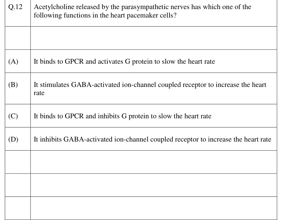
Acetylcholine released by the parasympathetic nerves has which one of the following functions in the heart pacemaker cells?
It binds to GPCR and activates G protein to slow the heart rate
It stimulates GABA-activated ion-channel coupled receptor to increase the heart rate
It binds to GPCR and inhibits G protein to slow the heart rate
It inhibits GABA-activated ion-channel coupled receptor to increase the heart rate
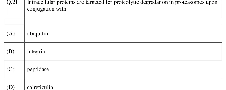
Intracellular proteins are targeted for proteolytic degradation in proteasomes upon conjugation with
ubiquitin
integrin
peptidase
calreticulin
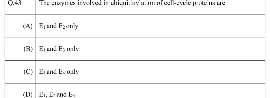
The enzymes involved in ubiquitinylation of cell-cycle proteins are
E₁ and E₂ only
E₁ and E₃ only
E₁ and E₄ only
E₁, E₂ and E₃
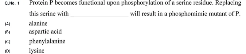
Protein P becomes functional upon phosphorylation of a serine residue. Replacing this serine with ________ will result in a phosphomimic mutant of P.
alanine
aspartic acid
phenylalanine
lysine
Ras protein is a
trimeric GTPase involved in relaying signal from cell surface to nucleus.
monomeric GTPase involved in relaying signal from cell surface to nucleus.
trimeric GTPase involved in regulation of cytoskeleton.
monomeric GTPase involved in regulation of cytoskeleton.
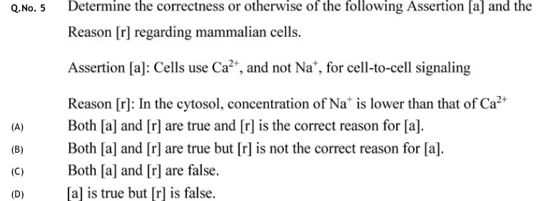
Determine the correctness or otherwise of the following Assertion [a] and the Reason [r] regarding mammalian cells.
Assertion [a]: Cells use , and not , for cell-to-cell signaling
Reason [r]: In the cytosol, concentration of is lower than that of
Both [a] and [r] are true and [r] is the correct reason for [a].
Both [a] and [r] are true but [r] is not the correct reason for [a].
Both [a] and [r] are false.
[a] is true but [r] is false.
Official answer: DSource: Official answer key, GATE 2020 BT (IIT Delhi)
PRACTICE
Which receptor class has intrinsic tyrosine kinase activity in its own cytoplasmic domain, activated when ligand binding brings two kinase domains together?
GPCR
Receptor tyrosine kinase (RTK)
Nuclear receptor
Ligand-gated ion channel
Answer: (B)
GPCRs (7-transmembrane) activate a separate heterotrimeric G protein and have no catalytic domain of their own.
Nuclear receptors are ligand-activated transcription factors, not kinases.
Ligand-gated ion channels gate ion flow directly and have no kinase activity.
Receptor tyrosine kinases carry their own kinase domain. Ligand binding brings two kinase domains together (by dimerization in most RTKs, or by a conformational change in the preformed insulin receptor dimer), and the domains cross-phosphorylate each other's tyrosines.
Which of the following statements about heterotrimeric G-protein signalling downstream of a GPCR are correct?
The GTP-bound form of Gα is the active state that signals to downstream effectors
Gα has intrinsic GTPase activity that terminates the signal by hydrolysing bound GTP
GPCRs contain an intracellular tyrosine kinase domain that is activated on ligand binding
A single activated GPCR can activate multiple G-protein molecules in succession, amplifying the signal
Answer: (A), (B), (D)
A is correct: GTP-bound is the active form that regulates downstream effectors.
B is correct: has intrinsic GTPase activity. Hydrolysing GTP to GDP ends its activity and allows it to reassociate with .
C is incorrect: an intracellular tyrosine kinase domain belongs to RTKs. A GPCR is a 7-transmembrane protein with no enzymatic domain; it activates a separate G protein.
D is correct: one ligand-bound GPCR can activate many G-protein molecules in turn before it is desensitised, which amplifies the signal.
A hormone binds its receptor at a single, non-cooperative site with . At a free hormone concentration of , calculate the percentage of receptors occupied by the hormone.
Answer: (accepted range )
Fractional occupancy for a single non-cooperative site: .
Substitute: .
As a percentage: .
A receptor binds its ligand with positive cooperativity and follows the Hill equation with Hill coefficient and . Calculate the percentage fractional saturation at .
Answer: (accepted range )
Compute .
Substitute: .
As a percentage: .
Check: half saturation is at . A non-cooperative receptor with the same would be only saturated at 20 nM, so cooperativity steepens the response, as expected.
Cholera toxin catalyses ADP-ribosylation of in intestinal epithelial cells. What is the direct consequence for cAMP signalling in these cells?
Gαs can no longer bind GTP, so cAMP falls
Gαs can no longer hydrolyse its bound GTP, so adenylyl cyclase stays active and cAMP stays high
Gαi can no longer be activated by its receptor, so inhibition of adenylyl cyclase is lost
cAMP phosphodiesterase is inhibited directly, so cAMP accumulates
Answer: (B)
ADP-ribosylation of blocks its intrinsic GTPase activity.
therefore stays in its GTP-bound (active) state and keeps stimulating adenylyl cyclase, so cAMP stays high.
A is wrong: GTP binding is not prevented; hydrolysis of the bound GTP is.
C describes pertussis toxin, which ADP-ribosylates . D is wrong: the toxin does not act on phosphodiesterase.
A ligand binds a -coupled receptor, activating phospholipase C-β (PLC-β), which cleaves membrane . Which pair of second messengers is generated directly by this reaction?
cAMP and PKA
IP3 and DAG
cGMP and PKG
Ca2+ only, with no lipid-derived messenger
Answer: (B)
-coupled receptors activate PLC-β, not adenylyl cyclase (adenylyl cyclase and cAMP belong to the / pathways).
PLC-β hydrolyses into two products: (inositol 1,4,5-trisphosphate) and DAG (diacylglycerol).
diffuses into the cytosol and opens -gated channels in the ER, releasing . DAG stays in the membrane and, together with , activates protein kinase C.
A (cAMP/PKA) is the pathway. C (cGMP/PKG) is made by guanylyl cyclases, e.g. soluble guanylyl cyclase activated by NO. D is wrong because is not a product of hydrolysis at all: it is released from the ER one step later, triggered by .
OFFICIAL GATE 2027 SYLLABUS WORDING
Copied from the GATE 2027 syllabus; see all 2027 syllabi.
GATE BT
Cell-cell communication; Cell signalling and signal transduction; Non-cell autonomous cell signalling; Post-translational modifications
Preparing in Kolkata? IAE Academy's course for GATE BT.