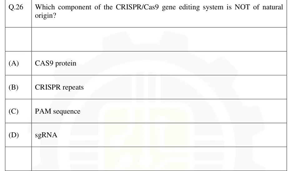
GATE 2026 BT Q.26
Which component of the CRISPR/Cas9 gene editing system is NOT of natural origin?
- (A)
CAS9 protein
- (B)
CRISPR repeats
- (C)
PAM sequence
- (D)
sgRNA

GATE BT
Notes cover site-directed mutagenesis (Kunkel, overlap-extension PCR, whole-plasmid PCR with DpnI, cassette), the CRISPR-Cas system (natural immunity, sgRNA, PAM, HNH/RuvC, NHEJ vs HDR, nickase, dCas9, base and prime editors, Cas12a, Cas13, off-target effects), biosensor components, transducers and performance (with the enzyme linear range, potentiometric slope and LOD convention), DNA-protein interaction tools (EMSA with fraction-bound and Kd, DNase I footprinting, ChIP, one-hybrid), protein-protein interaction tools (Y2H, Co-IP, pull-down, FRET, BiFC, SPR, AP-MS), and genomics/proteomics-scale approaches including ESI mass spectrometry, for GATE BT Recombinant DNA Technology.
3 previous-year questions · 3 solved · 6 practice questions
Site-directed mutagenesis is the deliberate introduction of a specific, pre-planned change (substitution, insertion or deletion) at a chosen position in a cloned DNA sequence, in order to study or alter the encoded protein's function, structure or regulatory behaviour. The exact base(s) changed are known in advance, which is what makes it useful for testing a specific hypothesis (e.g., "is this residue essential for catalysis?"). Random mutagenesis, by contrast, introduces changes at unpredictable positions (e.g., error-prone PCR, or chemical mutagens) and is followed by screening or selection for a phenotype.
Common methods
Oligonucleotide-directed (Kunkel) mutagenesis: a single-stranded, uracil-containing template is produced in a dut⁻ ung⁻ E. coli host (dut⁻ raises the dUTP pool so uracil is incorporated; ung⁻ stops it being removed). A synthetic mutagenic primer carrying the desired mismatch is annealed, extended in vitro by a non-strand-displacing DNA polymerase and sealed by DNA ligase. On transformation into a wild-type (ung⁺) host, uracil-DNA glycosylase removes the uracils from the parental strand, leaving abasic sites that inactivate it as a template, so the uracil-free mutant strand is strongly enriched.
PCR-based overlap-extension mutagenesis: two complementary internal primers, each carrying the desired mutation, are used with two flanking primers in two separate first-round PCRs to generate two fragments that overlap at the mutation. The fragments are mixed, denatured and re-annealed so their overlapping (mutant) ends prime each other, and a final PCR with only the flanking primers amplifies the full-length mutant product, which is then cloned.
Whole-plasmid PCR mutagenesis (single-tube, DpnI-based): a pair of complementary primers, each carrying the mutation, anneal at the same site on opposite strands of a double-stranded plasmid template and are extended around the whole plasmid by a high-fidelity, non-strand-displacing polymerase, giving nicked circular mutant plasmids. The parental template, isolated from a dam⁺ E. coli strain, carries methylated adenine in its GATC sites; DpnI cuts GATC only when the adenine is methylated (Gm6ATC), so it destroys the parental DNA, while the strands synthesised in vitro are unmethylated and survive. The nicks are sealed after transformation into E. coli.
Cassette mutagenesis: the wild-type restriction fragment spanning the site of interest is excised using two flanking restriction sites and replaced by a chemically synthesised double-stranded oligonucleotide 'cassette' carrying the desired mutation(s), then ligated back into the vector.
Comparing the methods
Method | Template needed | Key selection/enrichment step | Best suited for |
|---|---|---|---|
Kunkel (uracil-template) | Single-stranded, uracil-substituted (from a dut⁻ ung⁻ host) | ung⁺ host inactivates the uracil-containing parental strand | High mutant yield without needing a restriction site nearby |
Overlap-extension PCR | Double-stranded, any template | None needed: the whole mutant fragment is freshly amplified (then cloned) | Mutations far from a convenient restriction site; also insertions and deletions |
Whole-plasmid PCR (DpnI) | Double-stranded plasmid isolated from a dam⁺ strain | DpnI digestion of the methylated parental template | Fast, single-tube point mutations in a plasmid |
Cassette mutagenesis | Double-stranded, with two unique flanking restriction sites | Physical replacement of the fragment | Multiple or clustered mutations in one step |
Design and verification points
The mismatch is usually placed near the middle of the mutagenic primer, with enough correctly paired bases on each side for stable annealing.
PCR-based methods use a high-fidelity (proofreading) polymerase to limit unintended mutations elsewhere in the amplified DNA.
Every mutant clone is confirmed by sequencing the whole mutagenised region (or the whole insert), not just the target codon.
Alanine-scanning mutagenesis replaces residues one at a time with alanine to find which side chains a function depends on.
EXAM TIP
In Kunkel mutagenesis the enrichment works because the parental (wild-type) strand contains uracil and is inactivated in a ung⁺ host. It is not a restriction-based selection. In whole-plasmid PCR mutagenesis, the enrichment works because DpnI needs the GATC adenine methylated by E. coli Dam methylase. Plasmid prepared from a dam⁻ strain, or a PCR-amplified template, would defeat this step.
CRISPR-Cas is a bacterial and archaeal adaptive immune system repurposed as a programmable DNA-targeting tool. In the native system a CRISPR array (short repeats separated by spacers derived from earlier invaders) sits near the cas genes. Immunity runs in three stages. In adaptation, Cas1-Cas2 integrate a new spacer from invading DNA into the array. In crRNA biogenesis, the array is transcribed and processed into crRNAs; in type II systems a tracrRNA pairs with the repeats and RNase III processes them. In interference, the crRNA-guided effector cleaves matching invading nucleic acid. The PAM requirement lets the system distinguish invader DNA from its own array, where the spacer copies are not flanked by a PAM. Class 1 systems (types I, III, IV) use multi-subunit effector complexes. Class 2 systems (types II, V, VI) use a single effector protein: Cas9, Cas12 or Cas13.
For editing, a single guide RNA (sgRNA, an engineered fusion of crRNA and tracrRNA) directs the Cas9 endonuclease to a genomic site by Watson-Crick base pairing with a ~20-nucleotide target (protospacer) sequence, provided that the protospacer is immediately followed by a compatible PAM.
Core components and their roles
Guide RNA (sgRNA): a ~20-nt spacer complementary to the genomic target, fused to a scaffold that binds Cas9.
PAM (protospacer-adjacent motif): a short sequence immediately 3' of the protospacer on the non-target (PAM-containing) strand, recognised directly by the Cas9 protein, not by the guide RNA. For wild-type SpCas9 it is 5'-NGG-3' (N = any base); 5'-NAG-3' is recognised only weakly. Cleavage requires a compatible PAM, even when the guide matches the target perfectly.
Cas9 protein: an RNA-guided DNA endonuclease with two nuclease domains. HNH cleaves the target strand (the strand base-paired to the guide) and RuvC cleaves the non-target strand. Together they produce a blunt-ended double-strand break (DSB), typically 3 bp upstream (5') of the PAM.
Mechanism, in order
The sgRNA-Cas9 ribonucleoprotein complex scans DNA, pausing and locally unwinding wherever it finds a PAM.
If the adjacent sequence base-pairs with the guide spacer, starting from the PAM-proximal end, an R-loop forms and Cas9's nuclease domains are activated.
HNH and RuvC cut the target and non-target strands respectively, generating a blunt DSB.
The cell's own repair machinery resolves the break. NHEJ is error-prone and uses no template; it often introduces small indels that can disrupt the reading frame (the basis of gene knockout). HDR uses a donor template with homology arms and enables a precise correction or insertion, but it is largely restricted to S/G2 phase and is generally less efficient than NHEJ.
Cas variants and derived tools
Tool | What is changed | DNA cleavage | Typical use |
|---|---|---|---|
Wild-type SpCas9 | None | Blunt DSB ~3 bp upstream of NGG PAM | Knockout (via NHEJ), knock-in or correction (via HDR with a donor) |
Cas9 nickase (nCas9) | One nuclease domain inactivated: D10A (RuvC dead) or H840A (HNH dead) | Single-strand nick: D10A nicks the target strand, H840A the non-target strand | Paired nickases on opposite strands (a DSB only where both nick) for higher specificity; base and prime editors |
dCas9 (dead Cas9) | Both domains inactivated (D10A + H840A) | None; binds DNA only | CRISPRi (blocks transcription, or fused to a repressor domain), CRISPRa (fused to an activator domain), imaging |
Base editor | nCas9 or dCas9 fused to a deaminase | No DSB | Cytosine base editor: C•G → T•A; adenine base editor: A•T → G•C |
Prime editor | nCas9 (H840A) fused to a reverse transcriptase; the pegRNA carries the edit template | Nick only, no DSB, no donor DNA | Small substitutions, insertions and deletions |
Cas12a (Cpf1), type V | Different single-effector nuclease | Staggered DSB with 5' overhangs, distal to the PAM; T-rich PAM (e.g., 5'-TTTV-3') on the 5' side of the protospacer; needs only a crRNA (no tracrRNA) and processes its own crRNA array | Multiplexed editing; target-activated collateral ssDNA cleavage used in nucleic-acid detection |
Cas13, type VI | RNA-guided RNase (HEPN domains) | Does not cut DNA; cleaves target RNA | RNA knockdown; target-activated collateral RNA cleavage used in nucleic-acid detection |
EXAM TIP
Distinguish the two repair outcomes. NHEJ gives random small indels, needs no donor and operates throughout the cell cycle. An indel whose length is not a multiple of 3 shifts the downstream reading frame (knockout); a 3n-bp indel adds or removes whole codons in frame and may leave a partly functional protein, unless it creates a stop codon at the junction. HDR gives a precise, user-defined edit, needs a supplied donor with homology arms, is largely restricted to S/G2 and is less efficient. 'Insertion of a defined tag with homology arms' describes HDR; 'random frameshift knockout with no donor' describes NHEJ.
EXAM TIP
Off-target effects: mismatches between guide and DNA are tolerated more at the PAM-distal end than in the PAM-proximal 'seed' region (roughly the 10-12 nt next to the PAM). A genomic site with a few distal mismatches next to a PAM can therefore still be cut. Careful guide design, high-fidelity Cas9 variants and paired nickases reduce off-target cutting.
A biosensor is an analytical device that converts a biological recognition event into a measurable, usually electrical, signal. It has two essential functional parts, kept in direct contact with each other: a bioreceptor, which specifically recognises the analyte, and a transducer, which converts that recognition event into a quantifiable signal (current, potential, light, mass change or heat). Electronics then amplify and process the signal.
Classification by bioreceptor and by transducer
Classification basis | Type | Recognition/transduction principle |
|---|---|---|
Bioreceptor | Enzyme-based | Catalytic conversion of substrate produces a detectable product or consumes a co-substrate (e.g., glucose oxidase: glucose + O₂ → gluconolactone + H₂O₂) |
Bioreceptor | Immunosensor (antibody-based) | Highly specific antigen-antibody binding |
Bioreceptor | Nucleic-acid/aptamer-based (genosensor) | Sequence-specific hybridisation, or aptamer-target binding |
Bioreceptor | Whole-cell/microbial | Living cells respond to the analyte (e.g., via a reporter gene); can report bioavailability or toxicity, but response is generally slower and less selective than with a purified bioreceptor |
Transducer | Electrochemical (amperometric/potentiometric/conductometric) | Measures current, potential or conductance changes caused by the recognition event |
Transducer | Optical | Measures changes in absorbance, fluorescence, luminescence or refractive index (surface plasmon resonance) |
Transducer | Piezoelectric (mass-based) | A crystal's resonant frequency decreases when mass binds to its surface; for a thin rigid layer the decrease is approximately proportional to the added mass |
Transducer | Thermal (calorimetric) | Measures the heat released or absorbed by the binding or catalytic reaction |
General working sequence of a biosensor
The analyte in the sample diffuses to, and binds or reacts with, the immobilised bioreceptor.
The recognition event produces a physicochemical change (charge, mass, heat, light) at the transducer surface.
The transducer converts this change into an electrical signal.
The signal is amplified and processed by the associated electronics or software into a readable, usually quantitative, output.
Electrochemical transducers and the glucose biosensor
Amperometric: current measured at a fixed applied potential; the current is proportional to the rate of the electroactive reaction, and hence to analyte concentration within the linear range.
Potentiometric: potential measured at (near) zero current; it varies linearly with the logarithm of the ion activity (see formula).
Conductometric: change in solution conductance as the reaction produces or consumes ions.
Glucose sensor, first generation: glucose oxidase with measurement of O₂ consumed or H₂O₂ produced at the electrode (dependent on dissolved O₂).
Second generation: an artificial redox mediator shuttles electrons from the enzyme's reduced FAD to the electrode, reducing the O₂ dependence.
Third generation: direct electron transfer between the enzyme and the electrode, with no mediator.
WHY AN ENZYME BIOSENSOR RESPONDS LINEARLY AT LOW SUBSTRATE (MICHAELIS-MENTEN)
POTENTIOMETRIC RESPONSE (ION ACTIVITY $A_I$, ION CHARGE $Z_I$ WITH SIGN)
At 25 °C, mV, so the ideal slope is about mV per tenfold change in activity (about 59 mV per decade for a singly charged cation). Real sensors can be sub-ideal, so the slope is taken from the calibration.
Performance characteristics
Sensitivity: slope of the calibration curve (signal per unit concentration).
Selectivity: response to the analyte relative to interfering species.
Limit of detection (LOD): the lowest concentration distinguishable from the blank (see formula for a common convention).
Linear (dynamic) range, response time, stability (shelf and operational) and reproducibility.
LIMIT OF DETECTION, COMMON CONVENTION ($S_{\TEXT{BLANK}}$ = STANDARD DEVIATION OF BLANK SIGNAL, $S$ = CALIBRATION SLOPE)
EXAM TIP
By the usual definition, a biosensor has its bioreceptor integrated with the transducer and gives a direct signal. A conventional plate ELISA, whose colour is read afterwards in a separate plate reader, is an immunoassay rather than a biosensor, even though both rely on antibody-antigen recognition.
These techniques ask whether, and where, a protein (typically a transcription factor or another DNA-binding protein) contacts a DNA sequence. Some work in a test tube (in vitro); others work inside the cell, on native chromatin (in vivo).
EMSA (electrophoretic mobility shift assay, or gel-shift assay), in order
A short, labelled (radioactive or fluorescent) double-stranded DNA probe containing the putative binding site is incubated with the candidate protein (a purified protein, or a nuclear or cell extract).
The mixture is run on a native (non-denaturing) polyacrylamide gel alongside free probe alone.
Free DNA migrates fast. DNA bound by protein forms a larger complex that migrates more slowly and is seen as a retarded ('shifted') band.
Specificity is tested by competition: excess unlabelled probe of the same sequence (specific competitor) abolishes the shifted band, while an unrelated unlabelled DNA (non-specific competitor) does not.
Identity is tested with a specific antibody against the suspected protein. It either produces a further-retarded 'supershifted' band or, if it blocks the DNA-binding surface, abolishes the shifted band. Either result implicates that protein.
EMSA FRACTION BOUND ($I$ = BAND INTENSITY, SIGNAL PROPORTIONAL TO AMOUNT OF PROBE)
DISSOCIATION CONSTANT FOR 1:1 BINDING AT EQUILIBRIUM
When the labelled probe is at trace concentration (far below the protein), . At , . The method also assumes that the complex does not dissociate appreciably during electrophoresis.
DNase I footprinting, in order
A DNA fragment labelled at one end only (on one strand) is incubated with and without the DNA-binding protein.
Both samples are treated briefly with a limiting amount of DNase I, so that each molecule is cut on average no more than once (single-hit conditions) at a random accessible position.
The products are run on a high-resolution denaturing sequencing gel alongside a sequencing ladder of the same fragment (e.g., a G+A ladder).
In the protein-bound lane, the region physically covered by the protein is protected from cleavage and appears as a gap (the 'footprint') in the otherwise continuous ladder of bands. This maps the protected nucleotides. Enhanced (hypersensitive) bands sometimes flank the footprint where binding distorts the DNA. Smaller probes such as hydroxyl radicals, or base-modifying agents such as dimethyl sulfate (DMS), give finer contact maps.
Chromatin immunoprecipitation (ChIP), in order
Intact cells are treated with a cross-linking agent (commonly formaldehyde) to covalently fix protein-DNA contacts in place.
Chromatin is sheared (by sonication or nuclease digestion) into small fragments, typically about 200-1000 bp.
An antibody specific to the protein of interest immunoprecipitates the protein together with its cross-linked DNA. A non-specific IgG (or no-antibody) sample and an 'input' sample of sheared chromatin taken before immunoprecipitation serve as controls.
Cross-links are reversed and the DNA is purified. The recovered sequences are identified by PCR (or qPCR), microarray (ChIP-chip) or sequencing (ChIP-seq), revealing where the protein binds in the native genome.
Other DNA-protein tools
Yeast one-hybrid: a DNA sequence (the 'bait') is placed upstream of a reporter gene in yeast, and a library of proteins fused to a transcriptional activation domain is screened. Reporter expression identifies proteins that bind the bait sequence.
Southwestern blot: proteins separated by SDS-PAGE and blotted onto a membrane are probed with a labelled DNA sequence.
DNA-affinity chromatography: a binding-site DNA immobilised on a matrix purifies the proteins that bind it.
Reporter-gene assays: the effect of a protein, or of mutating a binding site, on transcription from a promoter-reporter construct tests the functional consequence of binding.
Comparing the three core techniques
Technique | Context | Resolution | What it establishes |
|---|---|---|---|
EMSA | In vitro | Low (just 'bound' vs 'free') | Whether binding occurs; with competitors and titration, specificity and apparent affinity |
DNase I footprinting | In vitro | Nucleotide level (the extent of the protected stretch) | The stretch of bases covered or protected by the protein |
ChIP | In vivo (native chromatin in intact cells) | Fragment level (hundreds of bp); refined by ChIP-seq peak calling and exonuclease-trimmed variants | Actual occupancy of a site inside the cell |
WORKED EXAMPLE
In an EMSA, a candidate protein is incubated with a labelled DNA probe. One lane shows a single shifted band. Adding a specific antibody against the candidate protein to a parallel reaction produces an even more slowly migrating band (a supershift), while an unrelated antibody produces no additional shift. What do these results establish?
The single shifted band (compared with free probe alone) shows that something in the reaction binds the DNA probe.
The specific antibody binds the protein already in the DNA-protein complex, adding mass and further retarding migration (supershift).
An unrelated antibody does not bind the complex and causes no additional shift, which rules out a nonspecific antibody effect.
Together, the results show that the candidate protein is present in the shifted complex. Sequence specificity of binding would additionally need specific and non-specific competitor lanes.
Answer: The supershift with the specific antibody, and its absence with the unrelated antibody, shows that the candidate protein is present in the DNA-protein complex responsible for the shift.
EXAM TIP
EMSA tells you whether binding happens. DNase I footprinting tells you where, down to which bases, it happens in vitro. ChIP tells you where it happens inside the cell, on native chromatin. Of these three techniques, only ChIP can confirm in vivo occupancy at native chromatin.
These techniques test whether two proteins (the 'bait' and the 'prey') physically interact. Some detect the interaction in a reconstituted reporter system in living cells (yeast two-hybrid, BiFC). Some co-isolate one protein with the other (co-immunoprecipitation, pull-down, affinity purification). Others measure proximity or binding physically (FRET, SPR).
Yeast two-hybrid (Y2H) assay, in order
Protein X (the 'bait') is fused to the DNA-binding domain (DBD) of a transcription factor, commonly GAL4. Protein Y (the 'prey') is fused to the activation domain (AD) of the same transcription factor.
Both fusion constructs are co-expressed in a yeast reporter strain carrying reporter genes (e.g., HIS3, lacZ) downstream of the transcription factor's binding site (UAS).
If X and Y interact, the DBD and AD are brought together, reconstituting a functional transcriptional activator.
This switches on the reporter genes, giving a detectable phenotype: growth on medium lacking histidine, or blue colonies on X-gal.
Y2H limitations
A bait that activates transcription on its own (autoactivation) gives false positives and must be tested alone first.
The interaction must occur in the yeast nucleus. Membrane proteins, or proteins needing modifications that yeast does not make, can give false negatives (split-ubiquitin variants are used for membrane proteins).
Yeast proteins can bridge the bait and prey, so a positive does not by itself prove direct binding.
Co-immunoprecipitation (Co-IP), in order
Cells expressing proteins X and Y are lysed under gentle, non-denaturing conditions that preserve native complexes.
An antibody specific to protein X is added to the lysate and captured on protein A/G beads.
The bead-antibody-X complex is washed to remove unbound material.
If Y was bound to X, it co-precipitates and is detected by Western blotting. This shows that X and Y associate in a cellular context, but it does not distinguish direct binding from binding bridged by another protein.
GST/His pull-down assay, in order
One purified, tagged protein (e.g., GST-tagged X) is immobilised on an affinity matrix: glutathione beads for GST, or Ni-NTA for a His tag.
Purified protein Y (untagged or differently tagged) is added and allowed to bind. GST alone on beads is run in parallel as the control.
Unbound protein is washed away.
Bound Y is eluted and detected by Western blot or staining. With only purified components and no cell lysate, retention of Y by GST-X but not by GST alone indicates direct binding. Caveats: a contaminant co-purifying with either protein could still bridge them, and proteins expressed in E. coli lack eukaryotic post-translational modifications.
Other protein-protein tools
FRET (Förster resonance energy transfer): a donor fluorophore on X transfers energy non-radiatively to an acceptor on Y only when they are very close (roughly 1-10 nm) and the donor's emission spectrum overlaps the acceptor's absorption. Efficiency falls with the sixth power of distance (see formula).
BiFC (bimolecular fluorescence complementation): two non-fluorescent halves of a fluorescent protein are fused to X and Y. Interaction brings the halves together and restores fluorescence in living cells (the reconstitution is essentially irreversible).
SPR (surface plasmon resonance): one partner is immobilised on a gold sensor surface and the other flows over it. Binding changes the refractive index near the surface, giving a label-free, real-time signal from which association and dissociation rate constants, and hence , are obtained.
Affinity purification-mass spectrometry (AP-MS), including tandem affinity purification (TAP) with two sequential purification steps under native conditions: identifies the members of a complex that co-purify with a tagged bait.
Chemical cross-linking followed by mass spectrometry: covalently links neighbouring proteins to capture transient or weak contacts.
FRET EFFICIENCY ($R$ = DONOR-ACCEPTOR DISTANCE, $R_0$ = DISTANCE AT WHICH EFFICIENCY IS 50%)
SPR DISSOCIATION CONSTANT ($K_{\TEXT{OFF}}$ IN S⁻¹, $K_{\TEXT{ON}}$ IN M⁻¹ S⁻¹, SO $K_D$ IN M)
WORKED EXAMPLE
A FRET pair has nm. Calculate the transfer efficiency when the donor-acceptor distance is (a) 2.5 nm and (b) 10 nm.
(a) , so .
.
(b) , so .
.
Answer: (a) ; (b) . Doubling the distance from cuts the efficiency from 0.5 to about 0.015, which is why FRET reports only very close proximity.
Comparing the core techniques
Technique | Context | Direct vs. possibly indirect | Notes |
|---|---|---|---|
Yeast two-hybrid | In vivo (yeast reporter system) | Possibly indirect (yeast proteins may bridge) | Suited to screening a library for new partners of a known bait |
Co-immunoprecipitation | Cell lysate (proteins expressed in cells) | Possibly indirect (another cellular protein may bridge) | Shows the association occurs in a cellular protein context |
GST/His pull-down | In vitro (purified components only) | Direct, if the purified proteins are free of bridging contaminants | Standard in vitro test for direct binding; run a tag-alone control |
FRET / BiFC | Living cells (or in vitro for FRET) | Proximity within a few nm | Reports where in the cell the interaction occurs |
SPR | In vitro, label-free | Direct (purified partners) | Gives , and |
Beyond single-gene or single-protein assays, whole-genome and whole-proteome methods characterise sequence, expression, interactions and function at system-wide scale. They are often combined with the targeted tools above: for example, ChIP followed by sequencing becomes ChIP-seq, a genome-wide map of one protein's binding sites.
Representative genomics-based approaches
Whole-genome sequencing (WGS): determines an organism's complete DNA sequence, either assembled de novo or by aligning reads to an existing reference genome, to identify variants and provide the coordinates on which other data are mapped.
RNA sequencing (RNA-seq): quantifies the transcriptome, revealing which genes are up- or down-regulated genome-wide after a mutation, treatment or CRISPR edit.
ChIP-seq: chromatin immunoprecipitation followed by next-generation sequencing, mapping genome-wide the sites a given DNA-binding protein occupies in vivo.
CRISPR-based functional screens: pooled libraries of guide RNAs knock out (or, with dCas9 fusions, repress or activate) genes across a cell population. A phenotypic readout (e.g., survival under a drug), followed by sequencing of the guides, identifies the genes that matter for that phenotype.
Representative proteomics-based approaches
Two-dimensional gel electrophoresis (2D-PAGE): separates a complex protein mixture first by isoelectric point (isoelectric focusing) and then by molecular mass (SDS-PAGE), resolving up to a few thousand proteins as spots for comparative expression analysis.
Mass spectrometry (MS): measures mass-to-charge ratio () of ionised molecules. MALDI (mostly singly charged ions) and ESI (multiply charged ions) are the common soft-ionisation methods. Proteins are identified by peptide mass fingerprinting (matching measured masses of digested peptides against a database) or by tandem MS (MS/MS) fragmentation, which reads peptide sequences, and post-translational modifications are located the same way. MS is the standard way to identify unknown partners recovered by Co-IP, pull-down or AP-MS.
Quantitative proteomics: stable-isotope metabolic labelling or isobaric chemical tags, or label-free comparison, measure relative protein amounts between samples.
Protein microarrays: thousands of purified proteins or antibodies spotted on a solid support to screen in parallel for binding partners, substrates or biomarkers.
Proteome-scale Y2H: one bait (or many baits) screened against an entire cDNA library to build a protein-protein interaction network (interactome).
ESI MASS SPECTROMETRY, PROTONATED IONS $[M+ZH]^{Z+}$ ($M$ IN DA, PROTON MASS ≈ 1.007 DA)
WORKED EXAMPLE
A protein of mass Da is analysed by ESI-MS in positive mode. At what does its ion appear?
For , the ion carries 15 extra protons: Da.
Ion mass Da.
.
Answer: . Multiple charging brings a large protein into the range of the analyser.
EXAM TIP
Each targeted technique in this topic has a genome- or proteome-scale counterpart. ChIP scales up to ChIP-seq, a single CRISPR knockout to a pooled CRISPR screen, a single Y2H test to a library screen or interactome, and Co-IP or pull-down to AP-MS with mass spectrometry. Learning them in these pairs makes it easy to see what each scaled-up method is measuring.
PREVIOUS-YEAR QUESTIONS
Try each question first, then open the answer. We placed each question under a topic ourselves; the exam paper does not label topics.
See these papers in full: GATE BT 2026 · GATE BT 2023 · GATE BT 2019
Which component of the CRISPR/Cas9 gene editing system is NOT of natural origin?
CAS9 protein
CRISPR repeats
PAM sequence
sgRNA
A protein has three identical sites arranged at the vertices of an equilateral triangle. If one site is filled with a dye (donor), the measured quantum yield () is 0.5. Filling one site with a donor dye and a second site with an acceptor dye results in of 0.25. The measured of one site filled with donor and the other two sites filled with acceptor dye (rounded off to three decimal places) is ____.
For site-directed mutagenesis, which one of the following restriction enzymes is used to digest methylated DNA?
KpnI
DpnI
XhoI
MluI
PRACTICE
For genome editing with Streptococcus pyogenes Cas9 (SpCas9), which protospacer-adjacent motif (PAM) must lie immediately 3' of the 20-nt protospacer, read on the PAM-containing (non-target) strand?
5'-NGG-3'
5'-TTTV-3'
5'-NAG-3' (exclusively)
5'-NNGRRT-3'
Answer: (A)
SpCas9 requires a PAM immediately 3' of the 20-nt protospacer on the strand not base-paired to the guide RNA.
For wild-type SpCas9 this PAM is 5'-NGG-3' (N = any base). NAG is recognised only weakly, so option C ('exclusively') is wrong.
5'-TTTV-3' is a T-rich Cas12a (Cpf1) PAM, which lies on the 5' side of the target, and 5'-NNGRRT-3' is the Staphylococcus aureus Cas9 (SaCas9) PAM. Both are distractors from other Cas proteins.
Hence the answer is A.
Which of the following statements about CRISPR-Cas9-mediated genome editing are correct?
Non-homologous end joining (NHEJ) repair of the Cas9-induced break is error-prone and typically produces small insertions or deletions (indels)
Homology-directed repair (HDR) requires a donor DNA template carrying homology arms flanking the desired edit
Cas9 uses its RuvC and HNH nuclease domains to cleave the two DNA strands and generate a blunt-ended double-strand break
The guide RNA base-pairs with the PAM sequence itself to direct Cas9 to the correct genomic site
Answer: (A), (B), (C)
A is correct: NHEJ is error-prone, needs no template, and its small indels are the basis of CRISPR knockouts by frameshift.
B is correct: a precise, user-designed edit (correction or knock-in) by HDR needs a supplied donor (a plasmid or a single-stranded oligonucleotide) whose homology arms flank the edit.
C is correct: HNH cuts the target (guide-paired) strand and RuvC cuts the non-target strand, giving a blunt double-strand break about 3 bp upstream of the PAM.
D is incorrect: the guide RNA pairs with the 20-nt protospacer, not the PAM. The PAM is recognised directly by the Cas9 protein.
Hence A, B and C are correct.
In an EMSA, a trace amount of radiolabelled DNA probe is incubated with a sequence-specific DNA-binding protein at 30 nM (the protein is in large excess over the probe, so the free protein concentration can be taken as 30 nM). After electrophoresis, the shifted (protein-bound) band gives 2500 units and the free-probe band gives 7500 units; band signal is proportional to the amount of probe. Assuming 1:1 binding at equilibrium and no dissociation of the complex during the run, what is the dissociation constant (in nM)?
Answer: (accepted range )
Fraction of probe bound: .
For 1:1 binding, , which rearranges to .
With nM: nM.
Check: , as measured.
A researcher wants to test whether purified recombinant protein X interacts directly with purified recombinant protein Y, with no other cellular protein present that could mediate an indirect (bridged) interaction. Which technique is MOST appropriate?
Yeast two-hybrid (Y2H) assay
Co-immunoprecipitation (Co-IP) from a whole-cell lysate
GST pull-down assay using purified GST-tagged X (immobilized on glutathione beads) mixed with purified untagged Y
Chromatin immunoprecipitation (ChIP) using an antibody against X
Answer: (C)
Y2H (A) takes place in living yeast cells, where endogenous yeast proteins could bridge an apparent interaction. It does not test the two purified proteins in isolation.
Co-IP (B) is done in a cell lysate containing thousands of other proteins, any of which could mediate an indirect interaction.
A GST pull-down (C) uses only the two purified proteins, with GST alone as the control, so it directly tests whether X binds Y with no third cellular protein present.
ChIP (D) detects protein-DNA association on chromatin, not a protein-protein interaction.
Hence the answer is C.
A donor-acceptor FRET pair has nm (the distance at which transfer efficiency is 50%). The measured FRET efficiency between donor-labelled protein X and acceptor-labelled protein Y is 0.20. Assuming a single fixed donor-acceptor distance, what is that distance in nm (to two decimal places)?
Answer: (accepted range )
, so .
.
.
nm.
Check: , so .
Which of the following statements correctly pair a technique with what it reveals about a DNA-protein or protein-protein interaction?
EMSA confirms that a protein binds somewhere on a given DNA fragment, but does not by itself map the exact nucleotides contacted
DNase I footprinting identifies the stretch of nucleotides protected from nuclease digestion by the bound protein
Chromatin immunoprecipitation (ChIP) detects protein-DNA binding at its native genomic location inside cells, using an antibody specific to the protein of interest
Yeast two-hybrid (Y2H) assay maps the exact DNA bases contacted by a DNA-binding protein
Answer: (A), (B), (C)
A is correct: an EMSA shift shows only that binding occurs (a slower-migrating band) and gives no base-level map of the contact site.
B is correct: the bound region is protected from DNase I cleavage and appears as a gap (footprint) in the cleavage ladder beside a sequencing ladder, mapping the protected nucleotides.
C is correct: ChIP cross-links proteins to DNA in intact cells with formaldehyde, immunoprecipitates the protein with a specific antibody, and recovers the bound genomic DNA, reporting in vivo occupancy.
D is incorrect: Y2H reports a protein-protein interaction through reconstitution of a transcriptional activator's DBD and AD. It does not map DNA contacts.
Hence A, B and C are correct.
OFFICIAL GATE 2027 SYLLABUS WORDING
Copied from the GATE 2027 syllabus; see all 2027 syllabi.
GATE BT
Site-directed mutagenesis; CRISPR-Cas; Biosensing and biosensors; DNA-protein and protein-protein interaction tools; Genomics and proteomics-based approaches
Preparing in Kolkata? IAE Academy's course for GATE BT.