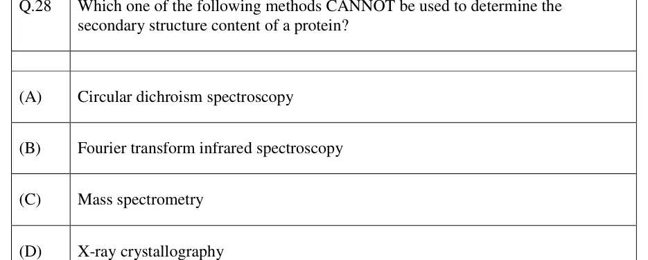
Which one of the following methods CANNOT be used to determine the secondary structure content of a protein?
- (A)
Circular dichroism spectroscopy
- (B)
Fourier transform infrared spectroscopy
- (C)
Mass spectrometry
- (D)
X-ray crystallography

Which one of the following methods CANNOT be used to determine the secondary structure content of a protein?
Circular dichroism spectroscopy
Fourier transform infrared spectroscopy
Mass spectrometry
X-ray crystallography
ANSWER
The correct answer is (C) Mass spectrometry.
Official answer: CSource: Official answer key, GATE 2023 BT (IIT Kanpur)
The question asks which technique CANNOT determine the secondary structure content (percentage α-helix, β-sheet, random coil) of a protein.
(A) Circular dichroism (CD) spectroscopy: the far-UV CD spectrum (190–250 nm) shows characteristic signatures for α-helix (double minima near 208 nm and 222 nm), β-sheet (minimum near 218 nm) and random coil, and is routinely deconvoluted to estimate % secondary structure — so CD CAN determine it.
(B) FTIR spectroscopy: the amide I band (about 1600–1700 cm⁻¹) position and its sub-band deconvolution directly reflect the fractions of α-helix, β-sheet, turns and unordered structure — so FTIR CAN determine it.
(D) X-ray crystallography: gives the atomic-resolution three-dimensional structure of the folded protein, from which secondary structure elements (helices, strands) are read out directly — so X-ray crystallography CAN determine it.
(C) Mass spectrometry: measures the mass-to-charge ratio of ionized peptides/proteins, giving molecular mass, amino-acid composition, sequence and post-translational modifications. It does not probe backbone dihedral angles or hydrogen-bonding patterns, so it CANNOT report secondary structure content.
Hence the technique that cannot determine secondary structure content is (C) Mass spectrometry.
KEY IDEA
Secondary structure content (%α-helix, %β-sheet, %random coil) of a protein is determined by spectroscopic/structural methods that probe backbone conformation and H-bonding — CD (far-UV signature bands), FTIR (amide I band deconvolution), and X-ray crystallography (atomic-resolution structure). Mass spectrometry measures mass-to-charge ratio and reports mass, composition, sequence and PTMs, not backbone conformation, so it cannot determine secondary structure content.
WATCH OUT FOR
Students sometimes assume any advanced structural/analytical technique (including MS) can report "structure" in a general sense, without distinguishing between primary-structure/mass information (MS) and conformational/secondary-structure information (CD, FTIR, X-ray).
From the GATE 2023 BT paper. Every question of this paper · Download the full paper · All 8 Microscopy & spectroscopy questions, with notes